In this article we will discuss about the external morphology of herdmania with help of suitable diagrams.
Shape, Size and Colouration:
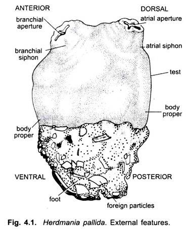
The body of Herdmania pallida is roughly oblong in outline, narrower at its attached than at its free end. At its free end, it is provided with two external openings—the branchial and atrial apertures. The average size of the adult is about 9.5 cm long, 7 cm broad and 4 cm thick. Older animals may even attain to a size of 12 x 8 x 4 cm, while an exceptionally large measures 13 x 8 x 4.5 cm. The foot, when present, may attain a length of as much as 3 to 4 cms. The general colour of the body in a fresh specimen is pink.
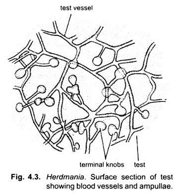
The presence of bright red patches, formed of terminal knobs (ampullae) in the blood vessels of the test, is a characteristic feature of Herdmania. The test is soft and leathery. It is more or less transparent in a young animal, but in an adult becomes usually opaque. The general surface of the test is much corrugated all over with lines, some shallow and others fairly deep, running in a criss-cross manner.
Body Divisions:
The body is divided into two parts, body proper and foot, and the entire animal is covered by the test.
ADVERTISEMENTS:
1. Body Proper:
It is the distal free portion of the body. The branchial aperture marks the anterior end of the animal; consequently the opposite end attached to the substratum is the posterior end. The side on which the atrial aperture is placed marks the dorsal side of the body, which is very limited. The side opposite to that on which the atrial opening is placed and which is partly attached to the substratum is ventral side which is extensive.
The branchial and atrial apertures are situated on short protuberances of the body called branchial and atrial siphons respectively. When fully extended, the atrial siphon is longer than the branchial. In a large-sized animal the atrial siphon measures about 1.5 cm, while the branchial siphon is only about 1 cm in length.
The atrial siphon is almost always directed upwards, the atrial aperture being more or less upright, while the branchial siphon is always bent a little outwards and the branchial aperture opens more or less laterally, always directed away from the atrial aperture. The average diameter of the branchial aperture, when fully expanded, is about 2 cm and that of the atrial aperture about 1.2 cm.
ADVERTISEMENTS:
At the base of branchial siphon, there is a ring of long branchial tentacles, while at the base of the atrial siphon there is a ring of slightly serrated folds which constitute the atrial tentacles. The test of the siphons is very elastic and can contract to close the apertures at the slightest disturbance of sea water.
2. Foot:
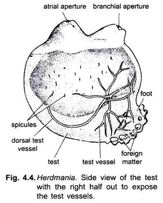
The foot, when present varies in character according to the nature of the substratum which the animal inhabits. If the substratum is of fine sand, the foot has an oval shape and a smooth surface and the test is quite hard in consistency. But if the substratum consists of coarse and the broken shell-pieces, the foot is irregular in outline and more or less soft in consistency.
Test or Tunic:
ADVERTISEMENTS:
The external covering surrounding the animal is a leathery, translucent test or tunic composed of tunicin, a substance akin to cellulose of plants. The foot is made entirely of test. It acts as a receptor as well as a respiratory organ. It is about 4-8 mm thick. It is ectodermal in origin.
The test has:
(1) A clear matrix in which is embedded,
(2) Cells of various shapes,
(3) Interlacing fibrils,
(4) Calcareous spicules, and
(5) Branching vascular vessels.
(1) Matrix:
It is gelatinous and made of a polysaccharide called tunicine.
ADVERTISEMENTS:
(2) Cells:
These are mesodermal in origin and have migrated into the test.
These are of 6 or 7 different types:
(a) Large eosinophilous cells,
(b) Small eosinophilous cells,
(c) Small amoeboid cells,
(d) Granular cells,
(e) Round vacuolated cells,
(f) Nerve cells with several processes, and
(g) Squamous epithelial cells.
(3) Interlacing fibrils:
These form a fine network in the test and resemble with smooth muscle fibres.
(4) Calcareous spicules:
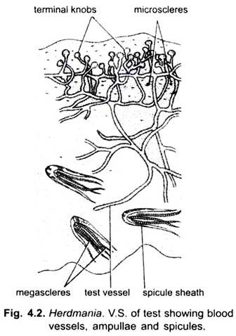
Herdmania has large number of calcareous spicules of two types. All the spicules bear several equidistant rings of minute spines all pointing in the same direction all along their length (Fig. 4.2).
(5) Blood vessels:
These blood vessels form a network in the test and end in bulb-like dilations called vascular ampullae. The vascular ampullae form vascular areas of bright red patches on the test. Vascular vessels and ampullae transport blood and, thus bring food to the test; they also act as accessory respiratory organs. The ampullae are also receptor organs.
Spicules:
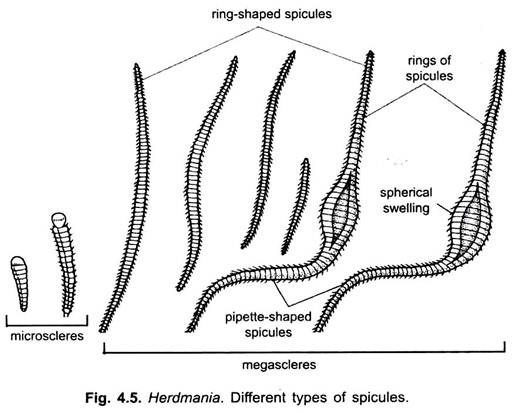
There are two types of spicules found in the body- small microscleres and large megascleres. They are all calcareous and have a definite shape.
1. Microscleres:
The microscleres are found only in the test and lie scattered throughout its substance. Each spicule consists of a rounded knob-like head and an elongated tapering body. The head is generally smooth, rarely with a few spines on its sides but the body bears a large number of rings of spines ranging from 5-20. These rings of spines run round the body of the spicule, nearly equidistant from one another. The spines always pointing towards the head of the spicule. The size of the spicule varies according to the stage of its growth, the average size being 50 microns, while very large ones measure about 80 microns.
2. Megascleres:
The megascleres are of two kinds:
(i) Spindle-shaped and
(ii) Pipette-shaped spicules.
Both these types of spicules are much larger than the microscleres.
(i) The spindle-shaped spicules are always found enclosed in a connective tissue sheath and lie scattered throughout the body of the animal in most of its tissues; but they are regularly arranged in the posterior half of the test. They are also very abundant in the mantle but are not uniformly distributed. They are usually very dense in the region of the stomach and the gonads and also at the bases of siphons, and are quite well represented in the region of the intestine but are sparse at the base of the longitudinal muscles.
They are usually arranged in linear rows in sheaths, each row having the appearance of a string. Like the microscleres, each spicule has a large number of rings of spines ranging from 20 to 60, all pointing in the same direction. Their size varies according to their stage of growth; an average size is 1.5 mm, but some attain a size of 2.5 mm.
(ii) The pipette-shaped spicules are larger than those of the spindle-shaped variety and sometimes assume immense proportions in size, being as much as 3.5 mm long. The characteristic feature of each spicule is the presence of a large spherical swelling in the middle which gives it the shape of a pipette when it is straight.
But very often the spicules are not straight, the two pointed limbs on either side of the swelling being bent one way or the other with the result the spicule assumes the shape of a U or V. They resemble the spindle-shaped spicules in having a large number of rings of spines and in possessing connective tissue sheaths. They are abundantly represented in the mantle, specially in the region of the gonads and lobes of the liver.
The megascleres are presents in almost every organ of the body. It is difficult to attribute any definite function to these spicules beyond saying that they probably form a skeletal tissue for the purpose of supporting the various organs of the body. It has been observed that a large number of spicules of the mantle project into the test, so that one-half of the spicule lies imbedded in the test and the other half in the mantle.
The function of some of these spicules, therefore, seems to keep the test firmly attached to the mantle. This view is confirmed by the fact that they are so situated the spines are all directed towards the mantle, so that mantle cannot be drawn away from the test when it contracts. Where the spicules form a sheath around the blood-vessels, their function appears to be stiffen the walls of the vessels and prevent their collapse.
Mantle or Body Wall:
Inside the test is the body wall or mantle. The mantle secretes the test. The mantle is present beneath the test and is attached only at the branchial and atrial apertures where it forms branchial and atrial siphons.
The mantle encloses a large atrial cavity or Prebranchial zone atrium, containing water. Histologically the mantle has an outer layer of ectoderm, middle layer of mesoderm, and an inner layer of endoderm forming the outer lining of atrial cavity.
1. Outer Ectoderm:
It is formed of a single layer of flat, hexagonal cells. It turns inside at pericardium the branchial and atrial apertures and extends up to the base of the siphons forming stomodaeum and proctodaeum respectively.
2. Inner Ectoderm:
It is formed of single layer of flat polygonal cells and forms the lining of the atrium.
3. Middle Mesoderm:
It lies beneath the outer ectoderm. It is composed of connective tissue traversed by muscle fibres, blood sinuses and nerve fibres.
The muscle fibres are of unstriped type and are arranged in three sets:
(i) Around the branchial and atrial siphons the mantle has annular muscles in several rings.
(ii) Below the annular muscles are longitudinal muscles which start from branchial and atrial apertures, then fan out up to the middle of the body on each side.
(iii) Branchioatrial muscles found in between two siphons.
