In this article we will discuss about the skeleton of rohu fish (Labeo rohita) with the help of suitable diagrams.
Exoskeleton:
Exoskeleton of Rohu Fish (Labeo rohita) comprises scales and fin-rays. Scales are cycloid type which are thin overlapping bony plates, partly covered by skin. Centrally each scale has an area called focus. Around focus there are concentric rows of ridges called circulii which are used to count the age of fishes.
Moreover, there are several radial lines, starting from focus and extending up to the periphery, called radii. Fin-rays are fin-supporting structures. They are of two types- spines (single ray) and soft rays (segmented rays). Spines in the fins of Labeo are unsegmented, uniserial structures. Soft rays are segmented, often branched and biserial.
Endoskeleton:
The endoskeleton of Labeo is completely ossified and forms the framework of the body. It is divided into an axial skeleton which includes the skull, vertebral column and ribs, and appendicular skeleton that includes the girdles and supporting elements of fins.
Skull:
ADVERTISEMENTS:
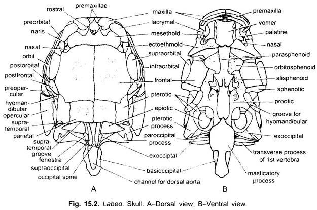
Skull of Labeo (Fig. 15.2) is an extremely complex structure, composed of many investing membrane or dermal bones and cartilage bones. The skull is divided into cranium enclosing the brain with paired olfactory, optic and auditory capsules for the respective sense organs and the visceral or branchial arches. The visceral arches are loosely attached with the skull.
1. Cranium and Sense Capsules:
The cranium and sense capsules are firmly united together.
ADVERTISEMENTS:
The cranium of Labeo rohita is divided into four regions:
(i) Ethmoidal region,
(ii) Orbitotemporal region,
(iii) Otic region,
ADVERTISEMENTS:
(iv) Occipital region.
(i) Ethmoidal Region:
It forms the anterior part of the cranium and is formed of paired nasals, ectoethmoids, lacrymals and unpaired median mesethmoid, vomer and rostral. The mesethmoid is a transversely placed bone, in front of the frontal bones. The rostral fits into a notch in the mesethmoid. The vomer is a quadrangular bone placed beneath the mesethmoid in front of the parasphenoid on the ventral side of the skull.
The nasals articulate with the posterolateral sides of the mesethmoid and form the roof of the olfactory capsules. The ectoethmoids also take part in the formation of olfactory capsules. Each has a deep nasal pit which lodges the nasal sac. The lacrymals are attached with the antero-lateral borders of mesethmoid on the antero-dorsal surface of the skull.
(ii) Orbito-Temporal Region:
In front of auditory region the cranium is excavated on each side by a large orbit. A vertical plate or inter-orbital septum separates the two cavities from one another. Anterior boundary of each orbit is formed by ectoethmoid, posterior boundary is formed by the sphenotic, dorsal boundary is made by frontal and ventral boundary is formed by alisphenoid and orbitosphenoid. Five small orbital bones form the orbital ring. These are anterior preorbital, posterior postorbital, supraorbital and postfrontal are of the dorsal side bones and infraorbital forms the anteroventral part of the ring.
Temporal region or sphenoidal region is further divided into:
(a) Anterior frontal region and
(b) Posterior parietal region.
ADVERTISEMENTS:
(a) Frontal region is formed of frontal bones on the dorsal side (also forms the roof of cranium). Orbitosphenoids and a median parasphenoid form the floor of this region. Frontals articulate anteriorly with the median mesethmoid and laterally and mesially with the nasals and ectoethmoids. Frontals articulate antero-laterally with the supraorbitals and postero-laterally with the postfrontals.
(b) Parietal region is formed of parietals on the dorsal side, alisphenoids form its lateral sides, and parasphenoid on the postero-ventral side. Basisphenoids are absent.
(iii) Otic Region:
Otic or auditory region is found behind the orbital region.
It is formed of:
(a) Anterior pro-otic which unites with its fellow of the opposite side in the floor of the brain case, just in front of the basioccipital;
(b) Opisthotic in the posterior part of the capsule, external to the exoccipital;
(c) Sphenotic above the pro-otic and forming part of the boundary of the orbit;
(d) Pterotic above the exoccipital and opisthotic, forming a distinct lateral ridge and produced behind into a prominent pterotic process; and
(e) Epitoic is a small bone wedged in between the supra- and exoccipitals and pterotic, and produced into a short epiotic process.
Opisthotic is absent in adult Rohu Fish (Labeo rohita). All these bones form an inverted cup-like structure that lodges the internal ear.
(iv) Occipital Region:
It forms the posterior most part of the skull. In the occipital region are four bones. These are the basioccipital, forming the greater part of the occipital condyle and the hinder region of the skull-floor, the paired exoccipitals placed one on each side of the foramen magnum and meeting both above and below it, and the supraoccipital, forming the occipital crest on the dorsal side. Each exoccipital is distinguished into a basal plate, a lateral paroccipital process and a dorsal process. Basioccipital is a long, tubular bone. It is thick in mid region.
2. Visceral Skeleton:
In fishes, it forms the part of the skull. It includes seven paired arches uniting in the mid-ventral line.
(i) The first or mandibular arch forms the jaws. It is formed of a palato-pterygoquadrate which forms the upper jaw and a Meckel’s cartilage that forms the lower jaw in the embryonic stage. Palatoquadrate is homologous with the upper jaw of dogfish. However, instead of remaining cartilaginous, it is ossified by three replacing bones. The toothed palatine lies in front, articulating with the olfactory capsule.
Metapterygoid projecting upwards from the quadrate, but between quadrate and the palatine are two investing bones, the ectopterygoid on the ventral, and the entopterygoid on the dorsal edge of the original cartilaginous bar.
The quadrate at the posterior end of the entopterygoid furnishes a condyle for the articulation of the lower jaw. These bones do not, however, enter into the gape, and do not form the actual upper jaw of the adult fish. External to them are two large investing bones, the premaxilla and the maxilla, which together form the actual or secondary upper jaw. Both these bones bear many teeth.
The lower jaw is formed of dentaries, angulars and articulars, Articular is formed by the ossification of Meckel’s cartilage and articulates with the quadrate of the upper jaw. Dentary meets with its fellow of the other side in the middle. All these bones bear no teeth.
(ii) The second or hyoid arch forms the suspensorium and hyoid cornu. Suspensorium is formed of two bones, the hyomandibular and symplectic. Hyomandibular articulates with the auditory capsule and on the other side with the symplectic. Hyoid cornu is composed of paired interhyal, epihyal, ceratohyal, hypohyal and a median unpaired basihyal. The epihyal and ceratohyal are provided with three sabre-shaped branchiostegal rays. Beneath the basihyal is present a median urohyal. Basihyal supports the tongue.
Operculum:
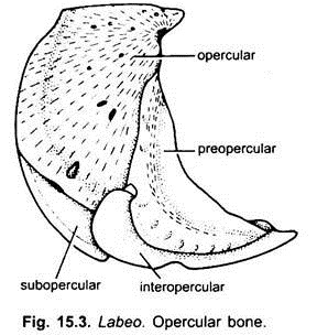
The operculum is formed of four investing bones; opercular, preopercular, subopercular and interopercular. The opercular is a large, flat bone with a condyle which articulates with the hyomandibular. The subopercular lies below and internal to the opercular.
The interopercular pits between the lower portions of the three preceding bones, and is attached by ligament to the angle of the mandible. The sabre-shaped branchiostegal rays are attached along the posterior border of the epi- and ceratohyal.
The remaining five arches are called branchial or gill-arches, diminishing in size from before backwards. First three branchial arches are composed of pharyngobranchial above, then epibranchial, then a large ceratobranchial, and a small hypobranchial below. The right and left hypobranchials of each arch are connected by an unpaired median basibranchial. All these segments are ossified by replacing bones.
The basibranchials are connected with one another and with the basihyal by cartilage so as to form a median ventral bar in the floor of the pharynx. In the fourth arch hypobranchial is absent.
The fifth arch is reduced to a single bone on each side. Small spine-like ossifications are attached in a single or double row along the inner aspect of each of the first four arches. These are the gill-rakers. They serve as a sieve to prevent the escape of food through the gill-slits and to protect the gill filaments.
Vertebral Column:
The vertebral column is differentiated into distinct bony vertebrae. It is divisible into an anterior or trunk region and a posterior caudal region.
1. Trunk Vertebra:
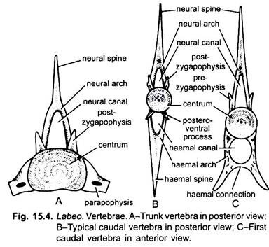
Trunk vertebrae are 21 in number. A typical trunk vertebra consists of a centrum with deeply concave anterior and posterior faces. In the embryonic stage the concavities are communicated by a narrow notochordal canal perforating the centrum. In the adult notochordal canal becomes closed. The low neural arches arise from the latero-dorsal surface of the centrum and fuse in the middle to form a long, slender neural spine directed upwards and backwards.
To the ventro-lateral region of the vertebra are attached by ligament a pair of long, slender pleural ribs with dilated heads. These curve downwards and backwards between the muscles and the peritoneum, thus, encircling the abdominal cavity. On either side of centrum is present the transverse process or parapophysis. On the anterior and posterior faces of neural arches are present pre- and postzygapophyses, by which adjacent vertebrae articulate.
The first four trunk vertebrae are greatly changed since these vertebrae connect the swim- bladder with the internal ear. In the last 3 or 4 trunk vertebrae the parapophyses are fused. The last vertebra also possesses the postero-ventral process.
2. Caudal Vertebrae:
The caudal vertebrae are 16 – 17 in number. In the caudal vertebrae the centrum is amphicoelous. The neural arches arising from the centrum are fused dorsally to form long, backwardly directed neural spine. Pre- and postzygapophyses are also present as in trunk vertebrae.
The outgrowths corresponding to the parapophyses are fused with the centrum and unite in the middle ventral line, forming a haemal arch. Through this run the caudal artery and vein. In the first six caudals each haemal arch bears a pair of ribs. In the rest the arch is produced downwards and backwards into a haemal spine.
The posterior end of the caudal region is enormously modified for the support of the tail fm. The hindmost centra have their axes deflected upwards and the last centrum is rod-like structure, the urostyle. The neural and haemal spines of the last five vertebrae are very broad and closely connected with one another.
They are more numerous than the centra, and three or four haemal arches are attached to the urostyle. In this way a firm vertical plate of bone is formed, to the edge of which the caudal fin rays are attached fanwise in a symmetrical manner.
3. Weberian Ossicle:
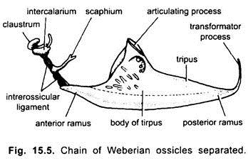
A more sensitive apparatus exists in the carps {e.g., Labeo) and siluroids (order Ostariophysi), in which a chain of bones connects the swim-bladder with the internal ear on either side, forming the Weberian apparatus. It is supposed that this apparatus is a modification of processes of certain vertebrae, which carries pressure stimuli anteriorly to the perilymph. It was discovered by Weber in 1820 who regarded them to be ear ossicles.
The first four trunk vertebrae have no parapophyses. The neural spine of the first vertebra is formed of two small pieces called claustrum and scaphium. These pieces are the two anterior most elements of Weberian apparatus. The centra of second and third vertebrae are completely fused to form a large centrum that carries a flattened triangular plate, the tripus.
Tripus is the modification of transverse process of third vertebra, and it is the last element of Weberian apparatus. In between scaphium and tripus is present a stout ligament. Embedded in this ligament is a small bone with a backwardly directed spinous process, which is called interclarium. It lies in between scaphium and tripus. Thus, there are four bones in the Weberian apparatus, viz., claustrum, scaphium, interclarium and tripus.
Skeleton of Median Fins:
The skeleton of median fins (dorsal and anal) consists of a series of:
(i) Somactidia or radials and
(ii) The dermotrichia or dermal fin rays.
Somactidia are parallel bony rods found embedded within the body muscles. Each somactid has proximal, mesial and distal segments. Dermotrichia support the fold of the fin. In Rohu fish (Labeo rohita), the dermotrichia or lepidotrichia which lie in the substance of the fin are slender bones, jointed together and mostly branched. At the free edges of fins are also present unbranched horny actinotrichia.
Dorsal fin is supported by 15 or 16 lepidotrichia placed on 14 somactidia. The proximal segment of each somactidia is large and dagger-shaped, and called axonast (interspinous bone). The mesial segment is short and distal segment is reduced.
Anal fin has a series of eight fin rays supported by seven somactidia.
Caudal fin is supported by a number of flattened bony rods. On the dorsal side of urostyle are present two epiurals and a radial, and on its ventral side are present nine hypiurals. The fin rays are attached with the epiurals and hypiurals in two symmetrical halves.
Appendicular Skeleton:
Pectoral Girdle and Fins:
1. Pectoral Girdle:
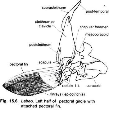
Each half of pectoral girdle (Fig. 15.6) is formed of:
(i) A scapula, situated dorsally to the glenoid facets and developed partly as a replacing, partly as an investing bone;
(ii) A coracoid situated ventrally to the glenoid facet and
(iii) A mesocoracoid situated above the coracoid and anterior to the scapula.
Externally to these is a very large investing bone, the cleithrum, extending downwards under the throat.
Its distal end is connected by means of a supraclavicle to a forked bone, the post temporal, one branch of which articulates with the epiotic and the other with the pterotic process. To the inner surface of the cleithrum are attached two flat scale-like bones with a slender rod-like postclavicle passing backwards and downwards among the muscles.
2. Pectoral Fin:
The pectoral fin is supported by nineteen lepidotrichia which are attached with four somactidia (radials). The radials articulate with the scapula.
Pelvic Girdle and Fins:
1. Pelvic Girdle:
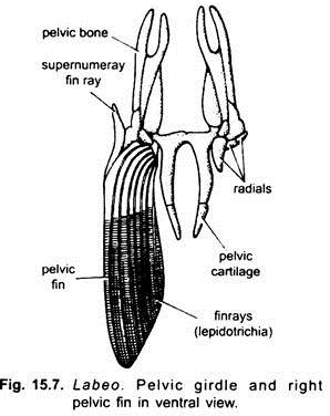
Pelvic girdle (Fig. 15.7) is placed anterior to anal fin, at the level of cloaca. Like pectoral girdle, it is also formed of two similar halves. Each half is formed of large, flat bone, the pelvic bone (basipterygium), lying in the middle line in the ventral body wall.
The pelvic bone has an anterior elongated part which is deeply forked at the tip, and a posterior rod-like cartilaginous part. The anterior forked end is connected with the rib of the twelfth trunk vertebra by ligaments. The posterior rod-like parts of both the halves are united in the mid-line.
2. Pelvic Fin:
Each pelvic fin is supported by nine lepidotrichia or fin rays and three somactidia or radials. The radials articulate with the posterior border of the pelvic bone. The first and second radials each bear two fin rays, and the third largest radial bears the remaining five fin-rays.
