In this article we will discuss about:- 1. Structure of Hormiphora 2. Enteron or Gastro Vascular System in Hormiphora 3. Histology 4. The Nervous System 5. Reproductive System 6. Development.
Contents:
- Structure of Hormiphora
- Enteron or Gastro Vascular System of Hormiphora
- Histology of Hormiphora
- The Nervous System of Hormiphora
- Reproductive System of Hormiphora
- Development of Hermiphora
1. Structure of Hormiphora:
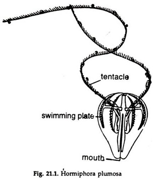
i. Hormiphora is pear-shaped, glossy, transparent, about 5-20 mm in diameter (Fig. 21.1).
ADVERTISEMENTS:
ii. From the opposite sides of the broad or aboral end, project two long, solid tentacles;
ADVERTISEMENTS:
a. The tentacles bear numerous lateral branches containing adhesive cells and
b. Are completely retractile into deep sheaths.
iii. Mouth, a slit-like opening present at the narrow oral end.
iv. Eight ad-radially arranged, equidistant meridional bands, the comb-ribs or costae or swimming plates, starting from near the aboral pole and extending about two-thirds of the distance towards the oral pole are present on the surface. These help in swimming.
ADVERTISEMENTS:
a. A row of transversely arranged comb-like structures of narrow plates, frayed at their outer ends constitute a comb-rib.
b. The combs are formed by the fusion of rows of cilia at their proximal ends.
v. An aboral sense organ is present in the depression at the centre of the aboral pole.
2. Enteron or Gastro Vascular System of Hormiphora:
a. The mouth leads to a flattened tube, the stomodaeum, which functions as stomach and extends to about two-thirds towards the aboral pole.
b. The walls of the stomodaeum are provided with internal ridges.
c. The stomodaeum narrows towards the aboral pole and opens in the infundibulum.
d. Running up to the aboral pole, the infundibulum divides into four short branches, two of which open to the exterior as minute excretory pores.
e. The other two branches of the infundibulum are par radial canals, pass outwards in the transverse plane and each divides into two adradial canals opening in the meridional canal.
f. Each par radial canal gives out a stomodeal canal and a tentacular canal.
3. Histology of Hormiphora:
ADVERTISEMENTS:
i. A delicate ectodermal epithelium constitutes the external covering of the animal. The cells forming the comb are large.
ii. The lining of the stomodaeum is ectodermal while that of the infundibulum and its canals is endodermal.
iii. A jelly-like mesoglea fills the spaces between the external ectoderm and the canal system.
iv. The tentacle sheath and the covering of the tentacles are ectodermal.
v. The longitudinal muscles constituting the core of the tentacles are of mesodermal origin.
vi. Delicate muscle fibres are present beneath the external epithelium covering the body and lining the canal system and the mesoglea.
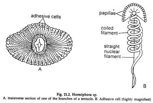
vii. The branches of the tentacles are covered by adhesive cells, which readily adhere the animal to an object (Fig. 21.2).
a. The outer surface of an adhesive cell is convex, bearing a number of papillae, which help to attach the tentacle firmly to an object.
b. A spirally coiled filament, with its delicate inner end extending to the muscular axis of the branch of the tentacle, is present inside the cell.
c. The filament acts as a spring to prevent the tearing of the adhesive cells, during struggle with the prey.
d. The nucleus of the cell is modified to a straight filament.
4. The Nervous System of Hormiphora:
The nervous system consists of sub-epithelial plexus of nerve fibres all over the body surface and constituted by multipolar ganglionic cells with their processes. The nerve fibres extend into the mesoglea.
Sense Organ in Hormiphora:
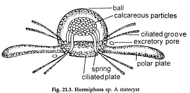
The principal sense organ is the aboral sense organ, which is a modified statocyst (Fig. 21.3) and help in maintaining equilibrium.
a. Two narrow areas of ciliated epithelium, the polar plates, are present in a depression at the centre of the aboral pole.
b. Four equidistant groups of very large S-shaped cilia, united to form as many springs, supporting a mass of calcareous particles, arise from the depression.
c. A pair of ciliated grooves from each spring run outwards and reach the swimming plates of the quadrant.
d. The calcareous mass with its springs is enclosed in a transparent case, formed of coalesced cilia.
6. Reproductive System of Hormiphora:
Hormiphora is hermaphrodite, i.e., testes and ovaries are present in the same individual.
a. Gonads are endodermal in origin and lie against the wall of the meridional canals.
b. The gonads are continuous or discontinuous. A testis extends along the whole length of the canal on one side, while the ovary extends along the whole length of the opposite side.
c. The gametes are discharged into the canals and escape to the exterior.
d. Fertilization is external.
7. Development of Hermiphora:
Development of Hormiphora is not very clearly understood.
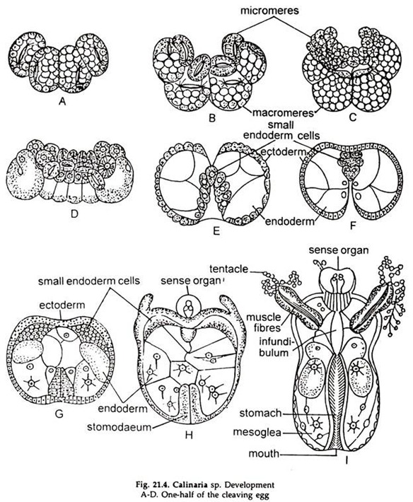
i. The zygote undergoes cleavage and eight micromeres and eight macromeres are formed (Fig. 21.4).
ii. The micromeres give rise to ectoderm and the macromeres are the source of endoderm.
iii. The micromeres divide rapidly, overspreads the macromeres, producing an embryo, having a central mass of large cells with yolk, partly surrounded by a layer of small cells, incomplete below.
iv. The endoderm cells multiply, invagination starts and a gastrula formed by epiboly and emboly, without an archenteron.
v. Stomodaeum is formed by invagination of the ectoderm, while the gastro vascular cavities arise by rapid multiplication of endoderm cells.
vi. The gelatinous mesoglea appears between the ectoderm and endoderm. The cells of the mesoglea are ectodermal in origin.
vii. The complex canal system of the adult appears.
viii. The swimming plates develop from bands of rapidly dividing ectoderm cells.
ix. The tentacles and their sheaths are derived from ectodermal thickenings on each side.
x. The muscle fibres and the calcareous bodies of the sense organs develop and the young hormiphora is formed.


