In this article we will discuss about:- 1. Pluteus Larva 2. Auricularia and Doliolaria Larvae 3. Doliolaria Larva of Crinoidea 4. Dipleurula Theory 5. Pentactula Theory 6. Regeneration in Echinoderms.
Pluteus Larva:
1. Five to six pairs of arms supported by calcareous rods and with pigmented tips.
2. Presence of four ciliated bands forming epaulettes at the base of the postoral and posterodorsal arms.
3. Arms are preoral, anterolateral, anterodorsal, postoral, posterodorsal and posterolateral.
ADVERTISEMENTS:
Two types of pluteus larvae, ophiopluteus and echinopluteus, are present:
Ophiopluteus larva:
1. Free-swimming.
2. Arms are four pairs, slender and supported by calcareous skeleton.
ADVERTISEMENTS:
3. Posterolateral arms are longest and directed forward, giving the larva V-shaped appearance.
4. Ciliated bands are present on the edges of arms.
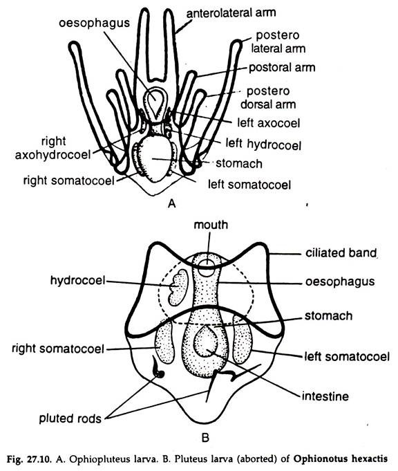
5. The alimentary canal is divisible into mouth, oesophagus, stomach and intestine opening through the anus (Fig. 27.10A).
Ophiopluteus is the larva of Ophiuroidea. In viviparous forms, Amphiura vivipara, the pluteus stage is omitted. In Ophionotus hexactis, the development takes place in ovary and pluteus larva (Fig. 27.10B) is devoid of arms and anus.
Echinopluteus larva:
1. Free-swimming.
2. Arms five or six pairs, pigmented and supported by calcareous skeleton.
3. The posterolateral arms are very short and directed outward or backward.
4. The skeletal rods simple or thorny or fenestrated or branched.
5. The zones of the alimentary canal are mouth, oesophagus, stomach and intestine opening through the anus.
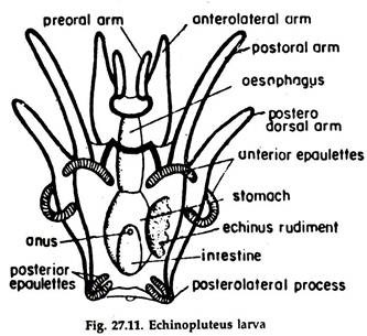
Echinopluteus (Fig. 27.11) is the larva of Echinoidea.
Auricularia and Doliolaria Larvae:
These larval forms occur in Holothuroidea.
ADVERTISEMENTS:
Auricularia larva:
1. A free-swimming form.
2. Body barrel-shaped and bilaterally symmetrical.
3. The preoral lobe is well formed.
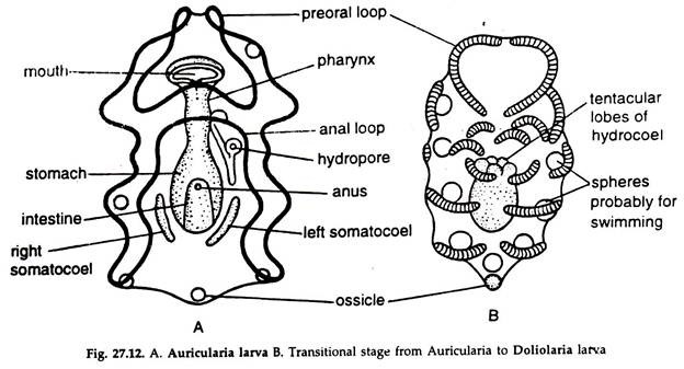
4. A single winding ciliated band, which may be produced into lobes (Fig. 27.12 A).
5. Gut with mouth, sacciform stomach, hydrocoel and right and left stomocoels and anus.
6. The hydrocoel becomes lobulated forming primary tentacles and communicates with the hydropore by a canal.
7. Calcareous rods replaced by spheroid or star-shaped or wheel-like bodies.
The auricularia larva is transformed into a Doliolaria larva similar to that of Crinoidea.
Doliolaria larva:
1. A free-swimming form.
2. Body barrel-shaped and bilaterally symmetrical.
3. Preoral lobe well-developed.
4. Wavy, continuous band break into 3-5 flagellated, transverse rings (Fig. 27.12B).
5. The gut with distinct zones.
Phylogenetic relationship:
Due to presence of enterocoelic coelom and some other minor resemblances, attempts have been made to establish a relationship between auricularia and tornaria larva of Hemichordata. This theory is now in dispute. Garstang (1894) propounded that the tadpole larva of Ascidia probably evolved from auricularia larva.
Doliolaria Larva of Crinoidea:
1. A free-swimming form.
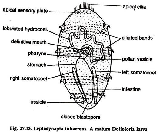
2. Body elongate oval, a little narrower posteriorly (Fig. 27.13).
3. Presence of 4-5 transverse ciliated bands around the body.
4. An apical sensory plate with a bunch of cilia at the anterior end.
5. An adhesive pit over the first ciliary band in the mid-ventral line close to the apical plate.
6. Gut with distinct zones and the stomodaeum between the second and third ciliated bands.
7. The skeletal structures present.
The larva attaches itself to some support and the internal organs rotate at 90° angle from ventral to posterior position. A stalk develops and the larva turns to a cystidian larva, which metamorphoses to a young individual.
Homology and phylogeny of echinoderm larvae:
Except for the crinoids, a sedentary group, the larvae of Asteroidea, Holothuroidea, Echinoidea and Ophiuroidea exhibit some fundamental resemblances.
1. Preoral and postoral loops.
2. Ciliated bands V-shaped.
3. Presence of gut with its divisions and openings.
4. Coelom enterocoelic.
These and other common features indicate they had a common ancestor.
Two theories have been forwarded to trace the common ancestry of different groups of echinoderms:
Dipleurula Theory:
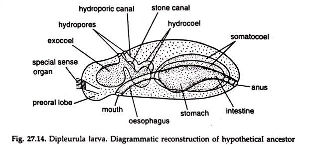
Bather (1900) suggests that different forms of echinoderms possibly evolved from a common ancestor resembling a hypothetical dipleurula larva.
Dipleurula larva:
1. Body oval, bilaterally symmetrical with a flat, ventral surface (Fig. 27.14).
2. Gut straight with a stomodaeum, stomach and proctodaeum. Anus is supposed to be formed by atriopore.
3. Three paired coelomic sacs—axocoel, hydrocoel and somatocoel, with water pores on the dorsal side.
4. Absence of skeletal structures.
The drawback of the concept is that it fails to explain the derivation of water vascular system, the most distinctive feature of echinoderms.
Pentactula Theory:
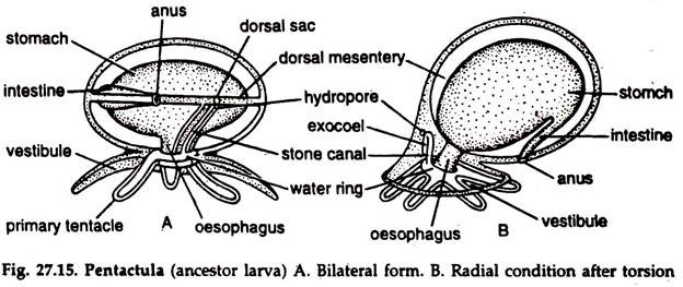
Advocated by Samon (1888), developed by Bury (1895), Hyman (1955). The concept advocates evolution of different groups of echinoderms from a common ancestor resembling pentactula larva. Pentactula larva.
1. Free, hollow tentacles around the mouth with coelomic branches from hydrocoel (Fig. 27.15).
2. Hydrocoel separated from the coelom to form water vascular system.
However, the opening of the system of coelomic canals on the surface through a canal developed from another portion of the coelom, i.e., the axocoel is not explained.
Divergence in larval forms follow pentacula stage:
a. The reproductive organs never became radial following their separation, the attachment was entirely lost during life history.
b. In asteroids and echinoids the gonads separated after the radial arrangement.
c. In crinoids the attachment of the gonads is retained.
It may be presumed that echinoderms are descendants of a common free-swimming ancestor, possibly pentactula.
Regeneration in Echinoderms:
Echinoderms have the remarkable ability to regenerate lost body parts following autotomy. In case, an arm is injured or grabbed by some predator or held up in some object, it is usually cast off (autotomy) near the base of the nearest ambulacral ossicle.
The broken end is immediately closed by the contraction of adjacent muscles of the body wall for protection of internal organs, and a new arm starts forming from the broken point.
