In this article we will discuss about:- 1. Description of Balanoglossus 2. External Features of Balanoglossus 3. Digestive System 4. Blood Vascular System 5. Nervous System 6. Reproductive System 7. Tornaria Larva 8. Metamorphosis.
Contents:
- Description of Balanoglossus
- External Features of Balanoglossus
- Digestive System of Balanoglossus
- Blood Vascular System of Balanoglossus
- Nervous System of Balanoglossus
- Reproductive System of Balanoglossus
- Tornaria Larva
- Metamorphosis of Balanoglossus
1. Description of Balanoglossus:
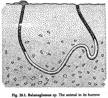
Balanoglossus belongs to class Enteropneusta, phylum Hemichordata. It is a marine animal, widely distributed in all seas of the world. It lives in burrows (Fig. 28.1) in inter-tidal zone and shallow water, though some species may live in somewhat deeper zones.
ADVERTISEMENTS:
The presence of Balanoglossus can be detected by the odour emitted by them, which resembles that of idoform. An individual lives in a U-shaped burrow excavated by the proboscis.
Sand grains adhere to a viscid matter secreted by numerous glands in the integument to form a fragile temporary tube. During low tide, the proboscis is protruded out of the burrow in search of food. The posterior end projects out through the other opening to eject faeces.
2. External Features of Balanoglossus:
ADVERTISEMENTS:
Balanoglossus is a soft-bodied, cylindrical, worm-like animal (Fig. 28.2). The size in different species varies from 2 to 3 mm to 2.5 metres. Most of the surface is ciliated.
The body is divisible into three regions— an anterior muscular, ovoid probosis:
The collar, just behind the proboscis; and a long, nearly cylindrical but somewhat depressed trunk.
ADVERTISEMENTS:
Proboscis:
The tongue-shaped proboscis has a thick, muscular wall, enclosing a cavity, the proboscis coelom, opening to the exterior by a minute proboscis pore. The narrow posterior or the stalk of the proboscis is strengthened by a layer of cartilage-like chon-droid tissue.
Collar:
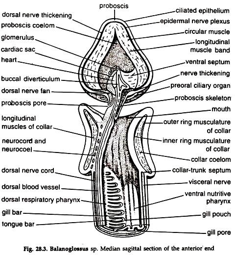
The collar is a prominent muscular fold encircling the proboscis stalk. It contains two coelomic cavities, the collar cavities, opening to the exterior by a pair of collar pores. The mouth (Fig. 28.3) lies on the ventral surface of the collar.
Trunk:
The trunk is long and constituted by branchiogenital region, hepatic region and caudal region. A double row of small pores, the branchial apertures, are present on the dorsal surface of the anterior end of the trunk, a row situated in a long furrow. During growth the apertures increase in number.
A pair of longitudinal genital ridges containing gonads are present in a considerable part of the trunk in the branchial region and behind. Two rows of prominences formed by the hepatic caeca constitute hepatic region. The rest of the trunk or the caudal region is nearly uniform in diameter and marked externally by irregular annulation.
Body wall:
ADVERTISEMENTS:
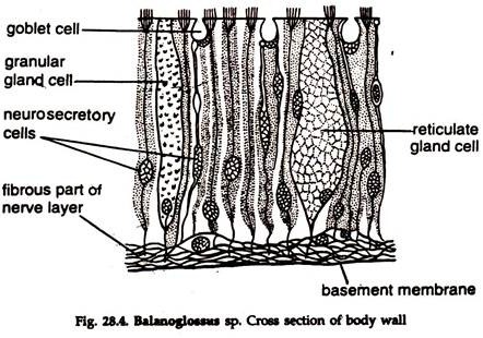
The outermost layer epidermis is made of tall and narrow ciliated cells. Three types of gland cells—reticulate gland cells, mulberry gland cells and goblet cells, are present in the epidermis.
A thick nervous layer lies beneath the epidermis, the two being separated by a thin layer of connective tissue, the muscular sheath (Fig. 28.4), supported by a basement membrane. The proboscis and collar contain circular muscle fibres, while longitudinal muscle fibres are present in the trunk region. The muscle fibres are smooth.
Notochord: buccal diverticulum; stomochord:
A diverticulum from the dorsal wall of the alimentary canal just behind the mouth, runs forward into the basal part of the proboscis, after sending out a small branch. The wall of the diverticulum is made of a single layer of very narrow, vacuolated cells of endodermal origin and encloses a narrow lumen.
The cells constituting the wall of the diverticulum are continuous with the epithelium of the alimentary canal (Fig. 28.3). This structure is considered as stomochord. The presence of stomochord in the anterior end of the body is possibly related to the animal’s principal mode of locomotion.
Proboscis skeleton:
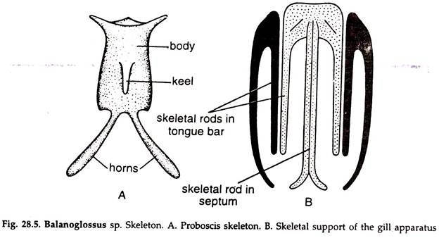
The proboscis skeleton is situated on the ventral surface of the buccal diverticulum, close to it. The skeleton consists of a hourglass shaped median plate with two back-wardly directed diverging limbs. A mid-ventral keel is present on the median plate (Fig. 28.5).
3. Digestive System of Balanoglossus:
The digestive system consists of a straight alimentary canal starting in mouth and ending in anus and divisible into pharynx, hepatic region and intestine. The whole of the alimentary canal is lined with cilia. The mouth is situated ventrally at the base of the proboscis, within the collar (Fig. 28.3).
The mouth leads into a small buccal tube, from the roof of which a buccal diverticulum protrudes into the proboscis coelom. The buccal tube opens into the pharynx. A peripharyngeal ridge on each side divides the pharyngeal cavity into two incomplete halves—dorsal and ventral (Fig. 28.6). The dorsal chamber, the repertory part, is pierced by gill slits and the ventral chamber is concerned with food collection.
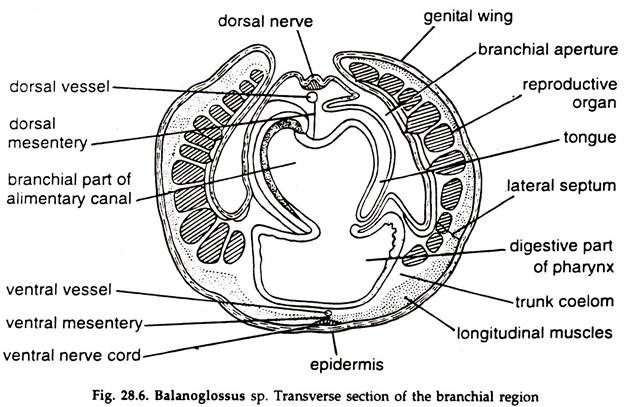 Internal gill slits open on the dorsal surface of the pharynx. Each gill slit is in the form of a long, narrow U, the two limbs separated by a narrow process, the tongue-bar containing a prolongation of the coelom. The internal gill slits lead into gill pouches, which, in turn, open to the exterior through gill pores.
The bars between the gill slits have skeletal supports, each of which is made of a dorsal basal portion and three long narrow rods. One rod is median and two are lateral. The median rod is bifurcated at the end and lies in the septum between the two adjacent gill sacs. The lateral rods lie in the adjoining tongue bars.
The pharynx opens posteriorly in the intestine. In the hepatic region, a series of paired hepatic caeca formed by the sacculation of the dorsal wall of the intestine are present. The post-hepatic region of the intestine is narrow and terminates in an anal aperture at the posterior extremity of the body.
Feeding of Balanoglossus:
Balanoglossus is a ciliary feeder. Cilia on the proboscis and lateral cilia on the sides of the gill bars play important role in feeding. Beating of the proboscis cilia drives food particles towards the mouth. A respiratory current is created by the beating of lateral cilia on the sides of the gill bars.
Water entering pharynx through the mouth is driven outwards through the branchial apertures. Food are swept directly into the mouth with respiratory current. Some food particles, silt and sand get entangled with the mucus secreted by the proboscis. The mucus, with its contents, is driven into the alimentary canal partly by the respiratory current and partly by the action of cilia in the buccal tube and gut.
Ciliary organ:
A ciliary organ bearing sensory cells, immediately anterior to the mouth, on the posteroventral surface of the proboscis is considered as an organ for testing water and matters drawn towards the mouth.
Digestion in Balanoglossus:
Nothing is definitely known about digestion in Balanoglossus. Amylase has been detected in proboscis. The oesophagus is most glandular and presumably secretes enzymes. The hepatic caeca secrete small amounts of maltase, proteinase and lipase.
4. Blood Vascular System of Balanoglossus:
A heart vesicle, blood vessels and sinuses constitute blood vascular system. Main longitudinal vessels are two; the dorsal vessel lies just below the dorsal nerve cord and the ventral vessel in the ventral mesentery. Blood flows forward in the dorsal vessel but backward in the ventral vessel.
The dorsal vessel runs forward from the region of anus to end anteriorly in a sinus, the dorsal sinus or heart, situated in the posterior part of the stomochord. A vessel arising from the posterior part of the sinus bifurcates to supply the proboscis. A number of vessels arising from the anterior part of the sinus forms a plexus, the glomerulus, possibly excretory in function.
An efferent vessel runs backward from each side of the posterior end of the glomerulus and breaks up into a plexus. The two plexus communicate with the ventral vessel and oxygenated blood is received by the ventral vessel.
The ventral vessel supplies the collar with a ventral collar vessel. Two distinct plexus in the collar tissue communicate with a ring vessel posteriorly. The ring vessel arises from the ventral vessel, located in the collar-trunk septum and it is connected with the dorsal vessel. The ventral vessel runs backward up to the anal region. It gives off lacunar networks along the whole length of the alimentary canal.
It sends a vessel to each gill septum, where it bifurcates to supply the two adjacent tongue bars. Each tongue bar receives two afferent branchial vessels, breaking up into a plexus. An efferent branchial vessel is formed from the plexus. The vessel runs dorsally, joins with the efferent branchial vessel of the adjacent tongue bar and opens into the dorsal vessel. Blood from the intestinal plexus is drained to the dorsal vessel.
A close cardiac sac encloses the dorsal surface of the heart. Muscle fibres present in the central wall of the sac contract rhythmically. Similar fibres are present in the dorsal and ventral vessels throughout the length of the trunk. Presumably all the muscles are responsible for driving blood in Balanoglossus.
5. Nervous System of Balanoglossus:
It consists of dorsal and ventral nerve strands extending throughout the length of the body, at the base of the epidermis. These are merely thickenings of a layer of nerve fibres. Giant nerve cells are scattered in the nerve strands. The nerve strands thicken to form two nerve cords, one in the mid-dorsal and the other in the mid-ventral line.
The two are connected by a peribranchial nerve ring in the collar- trunk septum. The dorsal cord projects into the collar coelom as the collar cord or neuro-cord- Nerve fibres run out from the ring. The dorsal and ventral nerve cords are conduction pathways.
Receptors and Sense Organs of Balanoglossus:
Preoral ciliary organ is the only organised receptor. Certain cells of the proboscis epidermis, on the anterior edge of the collar, might be sensory in nature.
6. Reproductive System of Balanoglossus:
The sexes are separate and often differ in shape and colour. Simple or branched saccular organs constitute testes or ovaries, arranged in a double row along the branchial region of the trunk and further behind. Each gonad opens to the exterior by a single pore.
Fertilization and development:
Fertilization is external. Development is associated with metamorphosis.
Early development:
Segmentation is complete, fairly regular and a round blastula is produced. Later, it becomes flattened. Gastrula is formed by invagination. At this stage the embryo is covered with short cilia and a ring of stronger cilia. Separation of ectoderm and endoderm is followed by hatching, which occurs at about 24-36 hours since fertilization.
The larva begins to swim. A diverticulum is separated from the anterior art of the archenteron. Proboscis coelom develops from the cavity of the diverticulum. Paired coelomic cavities of the collar and trunk develop either from the diverticulum of the posterior wall of the proboscis coelom or directly from the wall of the archenteron.
The free-swimming larva elongates and a transverse groove appears, posterior to the middle. Mouth appears by invagination in the position of the groove. Anus appears in the position of the blastopore. A second transverse groove divides the embryo into anterior, middle and posterior regions, the beginning of proboscis, collar and trunk. A tuft of apical cilia develop. The embryo is transformed into a Tornaria larva.
7. Tornaria Larva:
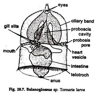
The size varies from 1 to 3 mm. Body bell-shaped, bilaterally symmetrical and variously folded (Fig. 28.7). The mouth is ventral and slightly posterior to the equatorial plane of the body. The anus is median and at the posterior end of the body. The portion anterior to the mouth forms the preoral lobe.
The anterior end bears an apical plate with nerve fibres, ganglionic nerve cells, a tuft of cilia and a pair of eye spots. Ciliated bands are three and distinct. The preoral and postoral bands join for a short distance at the apical plate.
The circumanal band surrounds the anus. The preoral and postoral bands collect food; the postoral and circumoral band or tolotroch are main locomotory organs. The alimentary canal is short and bent at right angle and divisible into oesophagus, stomach and intestine.
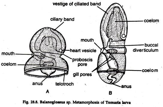
8. Metamorphosis of Balanoglossus:
Tornaria is pelagic. It sinks to bottom and undergoes a series of changes. The apical plate with its structures and ciliated bands are lost (Fig. 28.8). The preoral part of the body becomes ventrally ciliated and form proboscis, being separated from the collar by a constriction. Gill-slits begin to appear, trunk elongates and the larva assumes adult form.