In this article we will discuss about:- 1. Description of Lamellidens 2. External Features of Lamellidens 3. Microscopic Structure 4. Muscles 5. Locomotion 6. Body Cavity 7. Digestive System 8. Respiratory System 9. Circulatory System 10. Heart and Pericardium 11. Excretory System 12. Pericardial Gland 13. Nervous System 14. Receptors and Sense Organs in Lamellidens 15. Reproductive System 16. Fertilization and Development.
Contents:
- Description of Lamellidens
- External Features of Lamellidens
- Microscopic Structure of Lamellidens
- Muscles of Lamellidens
- Locomotion in Lamellidens
- Body Cavity of Lamellidens
- Digestive System of Lamellidens
- Respiratory System of Lamellidens
- Circulatory System of Lamellidens
- Heart and Pericardium
- Excretory System of Lamellidens
- Pericardial Gland of Lamellidens
- Nervous System of Lamellidens
- Receptors and Sense Organs in Lamellidens
- Reproductive System in Lamellidens
- Fertilization and Development in Lamellidens
1. Description of Lamellidens:
Lamellidens belongs to the class Pelecypoda phylum Mollusca. Fresh water mussels live in ponds, lakes, slow moving streams all over the world. The lamellidens are distributed throughout Indian subcontinent. Pearls of small sizes have been reported from lamellidens in West Bengal (India) and Bangladesh.
ADVERTISEMENTS:
The mussels remain partially embedded in mud or sand at the bottom of a water body. On slight disturbance, the foot and siphons are withdrawn inside the shell and the gape between the valves of the shell is tightly closed.
2. External Features of Lamellidens:
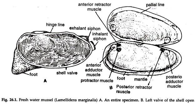
1. The mussel is enclosed in a hard, calcareous, horny-blackish, bivalve shell (Fig. 26.1A). The adult mussel is about 10 cm in length.
2. The body is light cream in colour, soft, elongate oval with a somewhat broad and a narrow posterior end.
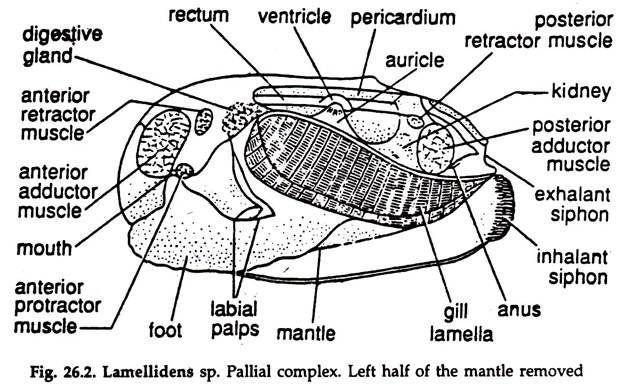
3. A manlte or pallium having two equal halves, the mantle lobes, lines the inner surface of the shell and encloses a space, the mantle or pallial cavity. The organs in the cavity are collectively termed pallial complex (Fig. 26.2).
ADVERTISEMENTS:
4. Two siphons—the dorsal exhalant siphon with smooth margin and the ventral inhalant siphon with fimbriated margin— formed by the mantle protrude beyond the shell at the posterior end.
5. In natural state, a narrow gape between the valves of the shell allows the muscular foot to project out from the anteroventral end of the body.
6. The viscerel mass, a pair of gills or ctenidia, two renal pores and the anus are lodged in the mantle cavity.
Shell:
ADVERTISEMENTS:
The shell consists of two equal halves or valves is made of conchiolin and calcic substances. It grows along the margins with the growth of the mussel.
1. The two valves are hinged along a Straight, dorsal hinge line by a tough, elastic hinge ligament, running transversely from one to another valve.
2. Each valve bears teeth-like projections, the hinge-teeth along the hinge line. The teeth of one valve fit with the sockets of the other.
3. Series of concentric rings, the lines of growth, widening towards the margin are present on the outer surface of the valve.
4. A small dorsomedian elevated region, the umbo is the thickest and oldest part of the shell. The lines of growth start from the umbo.
5. The inner surface of the shell is smooth, pearly in colour with a faint bluish tinge.
6. A narrow streak, the pallial line, marking the outer border of the mantle lobe is present on the inner surface of the shell, close to the margin.
7. Some scars, indicating the attachment of muscles are present on the inner surface of the shell (Fig. 26.1B):
a. Anterodorsal scars:
ADVERTISEMENTS:
i. Anterior adductor muscle—large, nearly round.
ii. Anterior retractor muscle—small, and posterodorsal to the adductor.
iii. Protractor muscle—small and posterior to the adductor.
b. Posterodorsal scars:
i. Posterior adductor muscle—large and oval.
ii. Posterior retractor muscle—small, close to the posterior adductor.
3. Microscopic Structure of Lamellidens:
The shell consists of three distinct layers:
i. Periostracum:
The thin outermost layer made of conchiolin, a substance related to chitin, and imparts colour to the shell.
ii. Ostracum or prismatic layer:
The thick middle layer, composed of alternate layers of conchiolin and prisms of calcic substances.
iii. Nacreous layer or mother of pearl:
The innermost layer composed of alternate layers of conchiolin and calcic substances. The layer is smooth and lustrous. The periostractum and ostracism are secreted by the mantle edge; soft and horny in colour at the beginning, which hardens with subsequent deposition of materials. The nacreous layer is secreted by the outer surface of the mantle. Pearl is secreted by this layer.
4. Muscles of Lamellidens:
The muscles are un-striated. The fibres of the muscles run from body to the shell. The foot muscles are not attached to the shell.
i. Adductor muscles:
Two, anterior and posterior: large transverse bands control the movements of the shell valves. Contraction closes the shells.
ii. Retractor muscles:
Two, anterior and posterior; run from the foot to the shell. Contraction causes retraction of the foot.
iii. Protractor muscles:
Two, anterior and posterior; contraction compresses the visceral mass.
iv. Pedal muscle:
The foot is made of a complex mass of intrinsic muscles.
5. Locomotion in Lamellidens:
The foot is the locomotors organ.
a. The foot is a muscular projection (Fig. 26.2) from the anteroventral end of the body and protrusible through the gape between the shell valves.
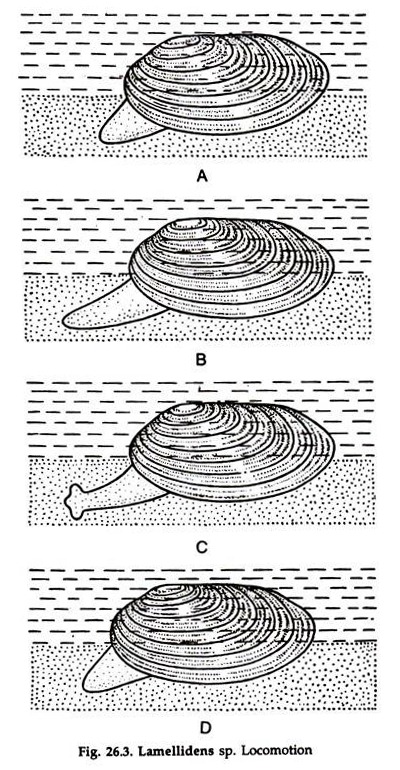
b. It is flattened from side to side and terminates in a more or less elongated keel, the anterior end being narrow, resembling a ploughshare (Fig. 26.3) which remains buried in the substratum.
c. A large sinus, the pedal sinus in the foot receives haemolymph from the pedal artery. Pressure on the haemolymph exerted by the contraction of foot muscles, forces the foot to extend forward.
d. In the next step, the narrow anterior end of the foot expands laterally and acts as an anchor in the substratum.
e. The foot muscles contract, the haemolymph from the sinus flows out through pedal vein and the mussel is drawn forward.
This is repeated and the mussel moves forward, ploughing its foot through the substratum.
6. Body Cavity of Lamellidens:
In the adult, the coelom is greatly reduced and restricted to pericardium (pericardial chamber), gonad (gonocoel) and lumen of the kidney (urocoel). The haemocoel acts as channels for haemolymph circulation.
7. Digestive System of Lamellidens:
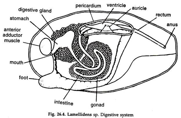
The digestive system consists of a coiled alimentary tract, two pairs of labial palps and a pair of digestive glands (Fig. 26.4):
Alimentary Canal:
Mouth:
Immediately below the anterior adductor muscles lies the mouth, which is a transverse slit. On each side of the mouth are two ciliated triangular flaps, the external and internal labial palps. The external palps unite in front of the mouth to form an upper lip and the internal palps unite behind the mouth to form a lower lip. The palps bear cilia.
Oesophagus:
A short tube, runs dorsally from the mouth to open into the stomach.
Stomach:
A large, oval sac embedded in the digestive gland. The epithelium is thrown into folds. Two narrow ducts from the digestive glands open in the stomach. Occasionally, a mucous rod, the crystalline style, which plays an important role in digestion is found in the stomach. It contains diastatic enzymes.
Intestine:
The intestine is a narrow tube and, after its origin from the ventral surface of the stomach near its posterior end, descends into the visceral mass above the foot, form two incomplete loops, ascends to the level of the stomach, anterior to the pericardium, turns sharply backward and proceeds straight as the rectum.
Rectum:
The rectum pierces the pericardium and the ventricle of the heart, runs parallel to the dorsal surface of the mussel and opens into the exhalant siphon by the anus. The internal wall of the rectum is thrown into longitudinal folds to form a ridge, known as typhlosole.
Digestive Glands:
The digestive glands are a pair of irregular light grey structures situated around the stomach. The secretion of the glands is discharged into the stomach through small ducts. Each gland is made up of numerous diverticula.
Feeding:
The fresh water mussel is a ciliary feeder, i. e. it collects food with the help of cilia. The food consists of detritus (partially decomposed organic particles) suspended in water, and microorganisms like bacteria, protozoa, etc. Detritus constitutes four-fifths of the total amount of food.
1. The respiratory water current created by the movement of cilia on the gills carries food particles and other matters.
2. The heavy particles drop in the mantle cavity and are expelled out through exhalant siphon by the movement of cilia in the mantle.
3. The gill laminae are covered with a thin sheet of mucus and the food particles are entangled in it.
4. The movement of lateral cilia on the gills drives mucous sheet to the ventral edge of the gills, bearing frontal cilia.
5. The frontal cilia drives the mucous string forward to the ciliary tracts of the labial palps.
6. Further assorting takes place here, the larger particles are carried to the tips of the labial palps, drop off in the mantle cavity and removed to the exterior.
7. The mucous string with the food matters are carried to the deep groove, the food groove between the two palps, leading to the mouth and driven to the oesophagus.
Digestion:
Not much is known about digestion in lamellidens. Intracellular digestion plays an important role in nutrition. Carbohydrates are only enzymes in the stomach and carbohydrate digestion takes place there. Intracellular digestion in the digestive glands hydropses protein and lipid to their constituents.
8. Respiratory System of Lamellidens:
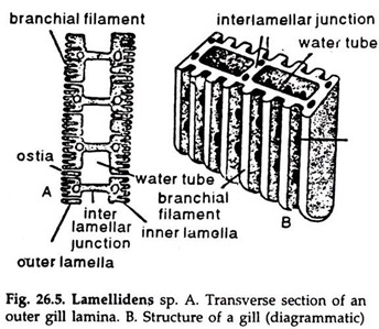
The respiratory organs consist of a pair of gills, one on each side between the body and the mantle (Fig. 26.2, 26.5):
1. Each gill consists of two laminae, the outer and the inner, and each lamina consists of two lamellae, outer and inner. The two lamellae of each lamina are united along the anterior, ventral and posterior edges, but free dorsally.
The cavity inside the lamina is divided into several chambers or water tubes by means of vertical interlameller junctions. Each lamella is made up of many gill filaments connected by interfilamentar junctions (Fig. 26.5). Minute pores or Ostia are present between the filaments.
2. The outer lamella of the outer lamina is attached to the mantle; the inner lamella of the outer and the outer lamella of the inner lamina are attached together to the visceral mass; the inner lamella of the inner lamina is attached to the visceral mass. All water tubes of the gill open dorsally into a supra-branchial chamber, which, in turn, opens to the exterior through the exhalant siphon.
3. The cells of the gill filaments are provided with cilia and, by their action, a current of water inhalant current, enters the mantle cavity through the inhalant siphon. From there it passes to the water lubes through Ostia and goes to the supra-branchial chamber to pass out through the exhalant siphon as exhalant water current. The gill filaments are highly vascular and gaseous exchange takes place there.
Heavier particles carried by the inhalant water current drop in the mantle cavity. Special cleansing cilia sweep small, non-food particles off the gill filaments. They are entangled with the mucus secreted by hypobranchial glands in the roof of the mantle cavity. The particles in the mantle cavity are swept to the exterior with exhalant water current.
4. The mantle is richly supplied with haemolymph and gaseous exchange occurs there also.
9. Circulatory System of Lamellidens:
A heart, arteries with their branches, sinuses and veins constitute, the open circulatory system of lamellidens. Leucocytes are few and suspended in the colourless haemolymph.
10. Heart and Pericardium:
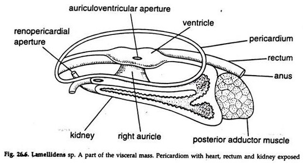
The heart consists of a single ventricle and two auricles, enclosed in a thin-walled pericardium (Fig. 26.6):
1. Ventricle:
A muscular chamber, surrounding the rectum.
2. Auricles:
Two thin-walled chambers, communicating with the ventricle by auriculoventricular apertures guarded by valves, opening only towards the ventricle.
Aortae:
An aorta arises from each end of the ventricle and from the point of origin they are named as anterior and posterior aorta:
1. Anterior aorta:
It arises from the anterior end of the ventricle and runs anteriorly above the rectum along the middle line.
a. At some distance from origin it bifurcates into two—the right and left terminal arteries.
i. Terminal artery:
Each sends branches to the mantle lobe, stomach, intestine, digestive gland, palp, foot and other organs at the anterior part of the body and finally terminates as the pallial artery.
2. Posterior aorta:
The aorta arises from the posterior end of the ventricle and runs posteriorly, ventral to the rectum, along the middle line.
The posterior aorta sends branches to the pericardium, heart, kidney, rectum and other organs at the posterior part of the body.
Sinuses:
The returning haemolymph from various parts of the body passes through many sinuses, smaller veins and finally collected in a large, longitudinal vena cava, lying between the kidneys.
Veins:
Haemolymph is returned to the heart through three circuits—mantle circuit, vena cava circuit and ctenidial circuit:
1. Mantle circuit:
Small veins arising from the mantle lobe join to form a large vein and returns oxygenated haemolymph directly to the auricle.
2. Vena cava circuit:
Prior to the origin of renal veins for supplying deoxygenated haemolymph to the kidney, a small vein arising from the vena cava, carries certain amount of deoxygenated haemolymph directly to the auricle.
3. Ctenidial circuit:
The major amount of haemolymph is returned to the auricle through this circuit.
a. Renal veins:
Carry deoxygenated haemolymph from vena cava to kidney.
b. Afferent branchial veins:
Carry haemolymph from kidney to the gills.
c. Efferent branchial veins:
Carry oxygenated haemolymph from gills to auricles.
11. Excretory System of Lamellidens:
A pair of kidneys or organs of Bojanus and a pericardial gland or keber’s organ constitute the excretory system.
Kidney:
1. The kidneys are more or less U-shaped tubes (Figs. 26.2, 26.6) located one on each side of the visceral mass, ventral to the pericardium.
2. The brownish, spongy, and glandular ventral arm is kidney proper, while the thin- walled, non-glandular, ciliated dorsal arm is the urinary bladder. The cilia produce an outward current.
3. The kidney proper opens in the pericardium through a Reno pericardial aperture and the bladder communicates anteriorly with its fellow of the other side by a large, oval aperture.
4. Each bladder opens in the mantle cavity through a minute renal aperture (nephridiopore) between the visceral mass and the inner gill lamina of the side.
12. Pericardial Gland of Lamellidens:
It is a reddish brown glandular mass, just in front of the pericardium and discharges the excretory matters in the pericardial cavity.
13. Nervous System of Lamellidens:
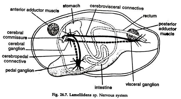
The nervous system consists of paired cerebral, pedal, visceral ganglia; commissure and connectives and nerves emanating from the ganglia and connectives (Fig. 26.7):
Cerebral ganglia:
Two, triangular in shape, yellow in colour, located at a distance from each other, lying at the base of the labial palps, just under the skin.
Cerebral Commissure:
A slender nerve, runs around the oesophagus in the anterior part of it and connects the two cerebral ganglia at their anterodorsal border.
Pedal ganglia:
The pedal ganglia are two, embedded deeply in the muscles of the foot at about 1 cm above the ventral border of the foot. The ganglia are oval in shape, pale yellow in colour and the two fuse medially to form a bilobed mass.
Cerbropedal connectives:
Arising from the posterior border of cerebral ganglia, the two nerves run downward, backward through the visceral mass and muscles of the foot to join the pedal ganglia at the dorsal border.
Visceral ganglia:
Two large ganglia, partially fused and located ventral to the posterior adductor muscle, covered by a thin peritoneum. The ganglia are flattened, triangular in shape
and pale yellow in colour. The two in vivo assumes X-shape.
Cerebrovisceral connectives:
Two, arising from the posterolateral border of cerebral ganglion, each runs backward, ventral to the attachment of the gill lamina with the visceral mass. It courses through the mass of the kidney and emerging from its posterior border runs further backwards to join the visceral ganglion of the side at its anterior border.
Innervation:
a. Nerves arising from cerebral ganglia innervate labial palps, anterior part of the mantle and adjacent structures.
b. Pedal ganglia send nerves to foot and anterior retractor muscle.
c. Visceral ganglia send following nerves.
i. Dorsal and posterior pallial nerves to the mantle lobes.
ii. Posterior renal nerves to kidneys.
iii. Branchial nerves to gills.
iv. Nerves to alimentary caneil.
Fine nerves from cerebropedal and cerebrovisceral connectives innervate the statocysts and structures in the visceral mass, respectively.
14. Receptors and Sense Organs in Lamellidens:
The sense organs are poorly developed. Paired osphradia, statocysts and scattered sensory cells constitute sense organs:
Osphradium:
A patch of brown pigmented sensory epithelium in association with each visceral ganglion. It is a chemoreceptor and tests the purity of the water entering mantle cavity.
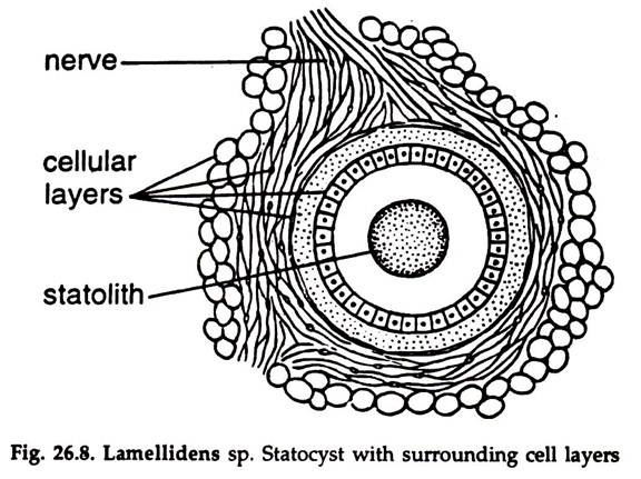
Statocyst:
The statocyst lies close to the pedal ganglion. It is a round sac, enclosing a space lined by a layer of sensory cells, supported by other cell layers (Fig. 26.8). A granule, the statolith is present in the space. The statocyst is innervated by the statocyst nerve arising from the cerebropedal connective. It is a balance organ.
Sensory cells occur on the outer epithelim of the body and inner border of the mantle. These act as tactile sense organs.
15. Reproductive System in Lamellidens:
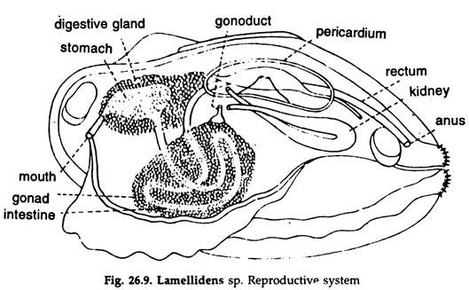
The sexes are separate; gonads are paired and occupy the major portion of the visceral mass in which the loops of the intestine are lodged. Both the testes and ovaries are racemose glands; the gonoducts arising from the gonads are short, narrowband open in the front of the renal aperture through a round genital pore (Fig. 26.9).
Male Reproductive System:
1. Testes white in colour.
2. A vas deferens arises from the posterolateral border of each testis.
Female Reproductive System:
1. Ovaries reddish in colour.
2. An oviduct arises from the posterolateral border of each ovary.
16. Fertilization and Development in Lamellidens:
Fertilization is internal.
1. The eggs discharged in the mantle cavity from the ovaries through female genital pores pass into the supra-branchial chamber.
2. The sperms discharged from the testes in the mantle cavity through male gonopores pass out with water current and reach the supra-branchial chamber of the female.
3. Fertilization takes place there and the fertilized eggs are passed to the water tubes.
4. In the water tubes, a number of fertilized eggs are enclosed in a thick, soft, somewhat triangular, whitish capsule, the egg capsule.
5. Development continues in the egg capsule and the capsules with glochidium larvae are discharged outside.
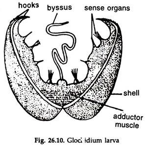
6. The glochidium larvae (Fig. 26.10) escape from the capsule.
Glochidium larva:
a. Presence of a bivalve shell.
b. Valves united dorsally and flat ventrally.
c. Ventral ends of the valves curve and bear spines.
d. Mantle lines the valves of the shell and bears brush-like hairs.
e. An adductor muscle at the base of the shell connects the valves.
f. Body is an undivided mass.
g. Presence of a byssus gland near adductor muscle.
h. A long byssus thread present.
Metamorphosis:
1. The glochidium larva attaches itself to the gill of a fresh water fish by the byssus thread.
2. The larva penetrates the host tissue, becomes enclosed by gill epithelium, live temporarily as ectoparasites and undergoes metamorphosis.
3. The byssus thread and sense organs disappear.
4. A stomodaeum is formed by ectodermal invagination, immediately posterior to the roof of the byssus thread and soon communicates with the archenteron. The opening of the stomodaeum forms the mouth.
5. Proctodaeum is not formed. The posterior end of the archenteron is in contact with the ectoderm and the anus is formed by a simple process of rupture.
6. Foot originates as a median, ventral elevation behind the mouth.
7. Gill rudiments appear as two papillae on each side of the mouth.
8. The larva fit for free life, drops from the host, sinks to the bottom and gradually attains adult form.
