In this article we will discuss about the circulatory system of pigeon.
Two different fluids circulate through the body of pigeon. One fluid, the blood, along with heart and the blood vessels constitute the blood vascular system. Another fluid, the lymph, and the lymph channels are included under the lymphatic system.
Blood-vascular system:
ADVERTISEMENTS:
This system includes blood, heart and the blood vessels:
(i) Blood:
Blood consists of plasma and corpuscles. The red blood corpuscles are oval in shape and nucleated. The white blood corpuscles are present in much lesser number, but are of different types.
The different types of white blood corpuscles are:
ADVERTISEMENTS:
(i) Lymphocytes,
(ii) Heterophils,
(iii) Polymorphonuclear-pseudo-eosinophilic granulocytes,
(iv) Basophils,
ADVERTISEMENTS:
(v) Eosinophils and
(vi) Monocytes.
Blood platelets are absent in pigeon, but the blood clots quickly. New blood cells are formed in the bone marrow and the blood corpuscles are destroyed within the spleen. Spleen is a red coloured body oval in shape, situated on the right side of the proventriculus and attached with it by peritoneum.
(ii) Heart:
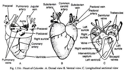
The heart is an oval organ placed in the anterior part of the thoracic cavity but ventral to the oesophagus. Heart is quite large in size in proportion to body size. It is enclosed by a thin white membranous pericardium and the pericardial cavity contains a serous fluid. The auricles and ventricles are distinctly separated by a groove called the coronary sulcus.
Sinus venosus is absent and is absorbed in the wall of the right auricle. Both auricle and ventricle are completely divided into right and left chambers (Fig. 1.116). Thus, heart is completely four- chambered and all the chambers are lined by endocardium.
The right auricle is slightly larger than the left auricle. The ventricles are very powerful. The left ventricle contains a round cavity while the right one has a crescentic cavity partly surrounding the left. The auricles and ventricles are divided internally by inter-auricular and inter-ventricular septa, respectively.
The right auriculoventricular valve is flap-like and muscular in nature. The presence of a single right auriculoventricular valve is a diagnostic feature of pigeon. The left auriculoventricular valve is membranous and provided with two cusps (bicuspids) which are attached with the ridges of the ventricular wall.
ADVERTISEMENTS:
Cord-like fibres (chordae tendineae) are attached to the margins of the auriculoventricular valves and to the walls of ventricles by papillary muscles. These muscles control the activity of the auriculoventricular valves via the chordae tendineae. The right auricle receives deoxygenated blood from three caval veins and the left auricle receives oxygenated blood through four pulmonary veins.
From the left ventricle the single right aortic arch originates and conveys oxygenated blood to the different parts of the body. The right ventricle gives rise to pulmonary arch which carries deoxygenated blood to the lungs. The left ventricle is usually called systemic ventricle and the right is called pulmonary ventricle.
The openings of the arches are guarded by three cup-like thick semilunar valves. The working of heart is controlled by elaborate intrinsic nervous system of heart. The wall of the right auricle bears sinuauricular node (or pacemaker) and the atrial septum bears auriculoventricular node. A special ring of Purkinje fibres is also present around the right auriculoventricular wall. The rate of systole and diastole is much faster than that in other vertebrates.
Mechanism of circulation through heart:
During the diastolic phase, the heart relaxes and the auricles receive blood from the veins. The right auricle gets the deoxygenated blood and the left auricle is filled up with oxygenated blood from the lungs via the pulmonary veins. The systolic action starts from the right auricle.
It actually begins from the sinuauricular node and passes to the auriculoventricular node. This wave then spreads to the remaining parts of the heart. At the time of auricular systole, the blood comes to ventricles through the auriculoventricular aperture. When the ventricles start contraction, the deoxygenated blood from the right ventricle is pushed to the lungs by the pulmonary arches.
Single right aortic arch from the left ventricle conveys the oxygenated blood to the different parts of the body. The heart of pigeon is a double circuit heart and there is no chance of mixing up of oxygenated and deoxygenated blood except in the capillaries. This is a significant evolutionary advancement in birds over reptiles.
(iii) Blood vessels:
The blood vessels include the arteries, veins and capillaries. The arteries supply blood to the different parts of the body and break up into arterioles and finally to finer anastomosing branches—the capillaries. The capillaries reunite to form the venules which ultimately form the veins.
Arterial system:
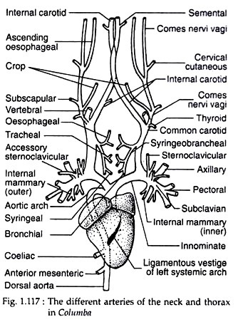
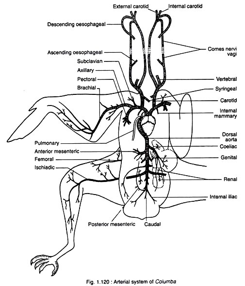
In pigeon, only the right aortic arch is present (Fig. 1.117 and 1.120). It arises from the left ventricle and passes backward between the auricles arching over the bronchus of the corresponding side. It then reaches the mid-dorsal line of dorsal body wall and runs backward as the dorsal aorta.
The innominate or brachiocephalic arteries are unequal in length; the right one is smaller than its left counterpart. The innominate arteries originate from the same region of the emergence of right aortic arch. The left systemic arch is absent in all adult birds. But vestige of the left systemic arch is present in the form of a solid ligamentous tissue extending obliquely forward (Fig. 1.117).
The arterial system of pigeon comprises of the following aortae and their branches:
Aortic arch:
An aortic arch originates from the left ventricle and then curves over the right bronchus. It reaches the dorsal body wall and then proceeds backwards as the dorsal aorta. This right aortic arch, immediately gives rise to two stout innominate or brachiocephalic arteries. Each innominate artery gives rise to common carotid and subclavian arteries. The common carotid arteries run parallel with each other along the neck region.
Each common carotid artery at the region of the thyroid gland divides into:
(a) A stout vertebral artery,
(b) A slender comes nervivagi, and
(c) An internal carotid artery.
The internal carotid arteries — after their emergence — converge anteromedially and run forward side by side through hypapophysial canal of the cervical vertebrae. In the anterior region of the neck the paired internal carotid arteries come out of the hypapophysial canal and depart laterally to give off external carotid arteries.
The other important arteries are:
A slender syringobronchial artery — supplies oesophagus, trachea, syrinx and bronchus. Comes nervi vagi artery—gives many branches to thyroid, crop, oesophagus, skin of neck etc. The comes nervi vagi artery passes alongside the vagus nerve and opens into the external carotid very near to its origin from the internal carotid artery.
A small anterolateral branch of comes nervi vagi gives off many smaller arteries. The external carotid artery gives origin to (i) hyomandibular artery, and (ii) facial artery. Both these arteries give off many tributaries (Fig. 1.117).
Subclavian artery:
The subclavian artery is a very stout vessel and gives rise to many arteries. After its origin it divides into
(i) an axillary artery, and
(ii) a pectoral artery.
Pectoral artery:
This artery branches profusely and supplies the breast muscles.
Axillary artery:
This artery is the continuation of the subclavian artery in the armpit or axilla. The axillary artery makes a slight curve and penetrates the brachial plexus and finally runs outward as the brachial artery to the arm. The pectoral artery ramifies into the pectoral muscles. The pectoral artery gives off a slender internal mammary artery (outer) which gives blood to the outer wall of the thoracic cavity.
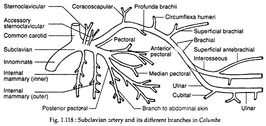
Some of the branches of the subclavian artery are (Fig. 1.118):
(i) Sternoclavicular artery gives branches to sternum, coracoid and clavicle.
(ii) Accessory sternoclavicular artery supplies blood to the adjacent muscles
(iii) Internal mammary artery (outer) supplies the inner wall of the chest cavity.
(iv) Axillary artery proceeds to the arm as the brachial artery and gives off (a) a coracoscapular branch (b) a profunda brachii, (c) a circumflexa humeri, and (d) a superficial brachial. Anteriorly, the axillary artery near the elbow-joint region divides into two unequal branches.
The branches are:
(i) Ulnar artery. This is a larger branch and gives cubital artery to the elbow joint and runs between the extensor and flexor muscles of the ulna.
(ii) Interosseous artery. It gives off a superficial ante-brachial artery into the pre-patagial muscle and proceeds anteriorly through the pronator muscles.
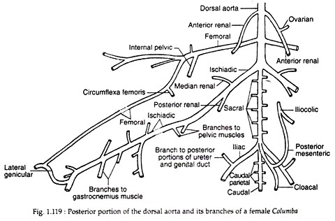
Dorsal aorta and its branches (Fig. 1.119):
The dorsal aorta runs along the mid-dorsal wall of the body cavity and sends the following branches:
(i) Dorsal intercostal artery:
It supplies the intercostal muscles.
(ii) Coeliac artery:
It arises from the dorsal aorta as a single artery to supply the abdominal viscera. It gives a short splenic artery to the spleen.
(iii) Anterior mesenteric artery:
It supplies the small intestine.
(iv) Genital artery:
This artery supplies to the gonad. In male, the testis gets the spermatic artery, while the female gets the ovarian artery to the ovary.
(v) Renal arteries:
The renal arteries comprise of three pairs of arteries supplying the three lobes of the kidney:
(a) Anterior renal arteries:
These paired arteries supply blood to the anterior lobe of the kidney,
(b) Median and posterior renal arteries:
Both these arteries are paired and supply the median and posterior lobes of the kidney.
(vi) Femoral artery:
These paired elongated branches pass through the kidney to supply blood to the proximal region of the hind limbs.
(vii) Ischiadic artery:
These paired arteries supply blood to the posterior part of the hind limbs.
(viii) Internal iliac artery:
The dorsal aorta divides posteriorly to form two internal iliac arteries, a posterior mesenteric artery and a single caudal artery.
(ix) Posterior mesenteric artery:
This single artery supplies the mesenteries of the posterior side.
(x) Caudal artery:
Single slender vessel originates as continuation of the dorsal aorta to supply the tail region.
Pulmonary arch:
The pulmonary arch arises from the right ventricle and immediately after coming out of the heart; it bifurcates to send pulmonary arteries to the lungs (Fig. 1.120). The pulmonary arch conveys deoxygenated blood from the heart to the lungs for oxygenation.
Venous system:
The venous system of pigeon is peculiar and shows the following characteristics:
(i) Each lung gives out two pulmonary veins opening into the left auricle.
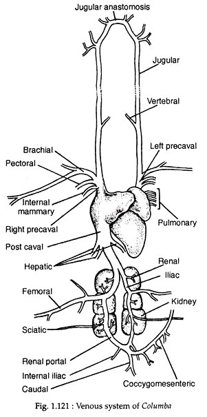
(ii) Two precavals and one postcaval open directly into the right auricle (Fig. 1.121). There is no trace of sinus venosus.
(iii) Considerable reduction of renal portal vein.
The veins in pigeon may be divided into three categories:
1. Pulmonary,
2. Systemic and
3. Portal veins.
1. Pulmonary veins:
The pulmonary veins constitute a very short circulatory circuit and carry oxygenated blood from the lungs. These veins enter the left auricle.
2. Systemic veins:
Three principal systemic veins — two precavals and one postcaval — drain deoxygenated blood from the capillaries of the body and open separately into the right auricle.
Veins anterior to the heart:
The paired precavals with all the veins opening into them are included under this category.
Each precaval receives:
(i) Jugular vein,
(ii) Brachial vein,
(iii) Pectoral vein, and
(iv) Internal mammary vein.
Jugular vein:
This vein receives several small veins from the crop and the shoulder, the vertebral vein and other veins from the head and neck. The vertebral vein brings blood from the vertebral column and spinal cord to the jugular vein. The veins from the crop and shoulder are small and numerous. Their number and disposition are variable—so they are not given specific names.
The left and right jugular veins are connected anteriorly by a small transverse connecting vein called jugular anastomosis. The anastomosis gets veins from the venous sinuses of the brain. This cross-connection in the jugular veins is a special adaptation for the flexibility of neck. The connection below the head prevents stoppage of blood circulation if one jugular vein is compressed during universal movement of the neck or head.
The jugular vein receives facial vein (carrying blood from the skin and muscles of the head), tracheal vein (brings blood from the trachea), cervical cutaneous vein (originates from a plexus in the skin of neck) and oesophageal vein (gets blood from the oesophagus). These small veins are not shown in Fig. 1.121.
The precaval vein is formed by the union of the following three veins:
Brachial vein:
The brachial vein receives blood from the corresponding wing. Some small branches from the shoulder also open into it.
Pectoral vein:
This vein is formed by the union of profusely branched veins from the pectoral region.
Internal mammary vein:
This vein brings blood from the sternum, coracoid region and the ribs.
Veins posterior to the heart:
The veins which are posterior to the heart include the following:
Postcaval vein:
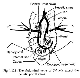
This vein (Fig. 1.121 and 1.122) is formed by the fusion of two iliac veins. Each iliac vein is the continuation of the femoral vein bringing blood from the leg region. The femoral vein passes through the kidney tissue. The postcaval receives few hepatic veins from the liver and a small vein from the ligament of the gizzard. Genital veins (spermatic vein in case of male and ovarian vein in female) are short veins which empty into the iliac veins.
Renal veins:
These veins bring blood from the kidneys and open into the iliacs as well as into the renal portal vein.
Sciatic vein:
This vein from the thigh opens into the renal portal vein.
Internal iliac veins:
These paired veins bring blood from the dorsal pelvic region.
Caudal vein:
This small vein comes from the uropodium. Coccygeomesenteric or inferior mesenteric vein. This vessel runs anteriorly in the mesentery t participate in the hepatic portal system. It also gets branches from the rectum. The blood from this vein also flows to the renal portal vein (Fig. 1.122).
3. Portal veins:
The hepatic and renal portal veins are also considered under the posterior veins. The renal portal vein originates at the junction of the coccygeomesenteric, internal iliac and caudal veins (Fig. 1.122). Each renal portal vein passes through the kidney tissue of that side and opens into the femoral vein and also receives sciatic vein.
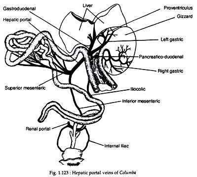
The renal portal vein is peculiar, because it never breaks up into capillaries in the kidney, but sends off a few small branches. Small renal veins open to this vessel. The hepatic portal vein forms an elaborated system. This system drains blood into the liver from the abdominal viscera (Fig. 1.123).
The hepatic portal system includes:
Gastro-duodenal vein which is formed by the pancreaticoduodenal vein and left gastric vein. The pancreaticoduodenal vein also gets a vein from the last part of the small intestine and the right gastric vein. The mesenteric veins are included under this system.
Lymphatic system:
The lymphatic system is well-developed and elaborate. Numerous lacteal vessels emerge from the small intestine. These vessels unite to form paired thoracic ducts. These ducts eventually open into the precaval veins.
