In this article we will discuss about the reproductive system of pigeon.
The sexes are separate. Sexual dimorphism is absent in pigeon.
Female reproductive system:
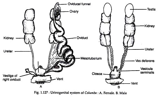
The female reproductive system is peculiar by having only left ovary and left oviduct (Fig. 1.127A).
ADVERTISEMENTS:
(i) Ovary:
The right ovary and oviduct are atrophied in adult. The left ovary is large and contains eggs of various sizes. The ovary is suspended from the dorsal body wall by a short mesentery called mesovarium.
(ii) Oviduct:
ADVERTISEMENTS:
The left oviduct is long and is attached with the dorsal body wall by a broad ligament or mesotubarium. The anterior end of the oviduct opens to the coelom by an expanded funnel-like opening called the oviducal funnel or ostium. The remaining part of the oviduct is thick, muscular and coiled. Various glands are present in the inner lining of the oviduct. When an ovum is matured, the ovarian follicle bursts to liberate the ovum.
The ovum enters into the cavity of the oviduct through the oviducal funnel. While passing down the oviduct, the ovum (fertilised or un-fertilised) becomes invested by the secretion of the various glands. The left oviduct opens into the urodaeum. A small vestigial right oviduct is found on the right side of the urodaeum.
Male reproductive system:
ADVERTISEMENTS:
The male reproductive system includes two testes and two vasa deferentia (Fig. 1.127B).
(i) Testes:
Each testis is an ovoid body which is attached to the anteroventral end of the kidney by a fold of peritoneum called mesorchium. The size of the testes varies greatly according to season. The testes are composed of numerous coiled seminiferous tubules. Between the tubules, groups of Leydig cells are present.
(ii) Vasa deferentia:
From the inner side of each testis originates a much coiled duct called vas deferens. Each vas deferens runs posteriorly, parallel with the ureter and opens into the urodaeum at the tip of a small papilla.
These papillae are slightly erectile and constitute the miniature copulatory organs of many birds. The last part of the vas deferens becomes slightly swollen to form seminal vesicle. Typical copulatory organs, observed in other vertebrates, are absent in pigeon.
Insemination and egg lying:
Fertilisation is internal. Insemination is done when the proctodaea of both the sexes are everted and brought close together in a state of ‘cloacal-kiss’. During this act, sperms are ejected into the female tract which travels up to fertilize the egg.
ADVERTISEMENTS:
Two eggs are generally laid at one time in pigeon. These eggs are incubated by the parents for a fortnight at a temperature of 38° to 40°C. When development becomes complete, young bird breaks the shell and comes out of the egg. They are nourished by the parents with pigeons milk.
Structure of egg:
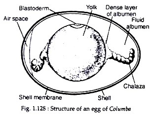
Egg is large in size due to accumulation of great quantity of yolk material. The protoplasm forms a small round area called germinal disc (blastodisc) containing the ovum. As it passes down the oviduct, a coat of thick albumen is accumulated and the disc becomes pushed to the upper side. As the egg rolls during its transit, the albumen becomes coiled at the two sides to form twisted cord—the chalaza.
In addition, more fluid albumen is deposited. Then a tough shell membrane and a calcareous shell are added by the secretory activities of the glands present in the oviduct. The shell membrane is parchment-like and is composed of two layers which enclose an air-space at the broader end of the egg (Fig. 1.128).
Shell is usually white in colour which may be coloured due to the deposition of special pigments. The shell consists of three layers and is provided with vertical pore canals.

