The following four points highlight the classification of protozoa:- 1. Entamoeba Histolytica Classification 2. Trypanosoma Classification 3. Leishmania Classification 4. Giardia Classification.
Type # 1. Entamoeba Histolytica Classification:
Phylum: Protozoa Microscopic and acellular
Subphylum: Plasmodroma Cilia absent; locomotion either by pseudopodia or flagella
Class: Rhizopoda Pseudopodia chief organs of locomotion and food catching.
ADVERTISEMENTS:
Order: Lobosa Pseudopodia short, blunt lobose type.
Sub-order: Nuda Body naked or enclosed in a thin membrane.
Genus: Entamoeba
Species: Histolytica
ADVERTISEMENTS:
Comments:
1. Entamoeba histolytica tropical and subtropical countries.
2. It occurs in the colon of man and feeds on the mucous membrane destroying the tissues by an enzyme which it secretes.
3. The size varies from 0.06 mm to 0.06 mm in diameter.
ADVERTISEMENTS:
4. It is dimorphic occurring in two forms:
(i)Trophic ortrophozoite,
(ii) Precystic or minuta.
Both these forms occur in man. Trophozoite is pathogenic, while minuta is non- pathogenic.
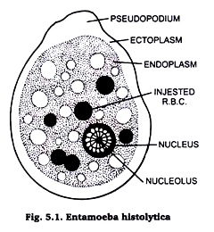
5. Cytoplasm is differentiated into ectoplasm and endoplasm.
6. Endoplasm contains single spherical nucleus and food vacuoles.
7. Contractile vacuole is absent.
8. One or two pseudopodia are present.
ADVERTISEMENTS:
9. Reproduction by binary fission and spore formation.
10. E. histolytica causes amoebic dysentery. When it infects liver, it causes amoebic hepatitis.
11. Mode of infection is oral through contaminated food or water with cysts of E. histolytica.
Habit & Habitat:
Entamoeba hystolytica is found as a parasite in the digestive tract of man, frog, and cockroaches in the tropical & subtropical regions.
Type # 2. Trypanosoma Classification:
Phylum: Protozoa Microscopic and acellular
Subphylum: Plasmodroma Cilia absent; locomotion either by pseudopodia or flagella
Class: Mastigophora Locomotory organs flagella.
Order: Protomonadina Amoeboid or euglenoid flagellates; flagella usually one or two; mouth and gullet absent; mostly parasitic.
Genus: Trypanosoma
Comments:
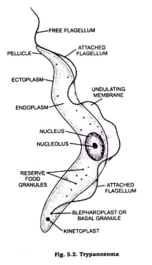
1. Trypanosome (Fig. 5.2) is polymorphic.
2. It has a colourless, slender, elongated, spindle-shaped body tapering at both ends, ranging in length from 15 to 16 micron.
3. Body is covered by the periplast or pellicle somewhat compressed laterally and twisted spirally.
4. The anterior end is more pointed than the posterior end and it bears the flagellum.
5. The body is convex on one edge and concave on the other, the convex edge of the body is thrown into irregular folds and is called undulating membrane.
6. Along the edge of the undulating membrane runs a stout thread or axoneme which is attached with the basal granule (blepheoplast) at the posterior end. Just posterior to the basal granule is a small rod-shaped body, the parabasal body (kinetoplast).
7. Nucleus is rounded and central in position.
8. Contractile vacuoles are absent.
9. Nutrition is saprozoic and all members are parasitic and pathogenic.
10. Reproduction takes place by longitudinal binary fission.
11. Most common species are as follows:
(i) T. gambiense parasitizes man in Central Africa causing sleeping sickness disease.
(ii) T. brucei parasitizes African cattle and causes fatal disease called Nagana.
(iii) T. cruzi causes Chagas disease which is restricted to South and Central America.
Habit and habitat:
Trypanosomes are found as parasites in the blood of vertebrates and are transmitted to them by the blood sucking invertebrates.
Type # 3. Leishmania Classification:
Phylum: Protozoa Characters same as those of Tryponosoma
Subphylum: Plasmodroma ………….–do-
Class: Mastigophora ………………….–do-
Order: Protomonadina Pseudopodia short, blunt lobose type.
Genus: Leishmania
Comments:
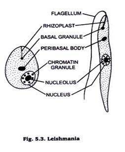
The genus Leishmania (Fig. 5.3) includes three species which are common parasites of man, viz:
(i) Leishmania donovani causing kala-azar,
(ii) Leishmani tropica causing oriental sore and
(iii) Leishmania brassiliensis causing American leishmaniasis.
Leishmania donovani has two phases in the life cycle, on flagellar Leishmania form in the peticulo-endothelial cells of man and the other Leishmania form in the gut of blood sucking fly phlebotomus. Leishmania form is intracellular and is found in the cells of liver, spleen, bone marrow, intestine and lymph glands in the reticulo-endothelial system.
This form is oval or spherical 1-3 µ in diameter with a limiting membrane, the periplast or pellicle. The cytoplasm contains an oval nucleus, rod-shaped or dot-kinetoplast and aparabasal body. Reproduction by binary fission.
Type # 4. Giardia Classification:
Phylum: Protozoa characters same as those of Tryponosoma
Subphylum: Plasmodroma…………. –do-
Class: Mastigophora ……………. –do-
Order: Polymastigina pseudopodia short, blunt lobose type.
Genus: Giardia
Comments:
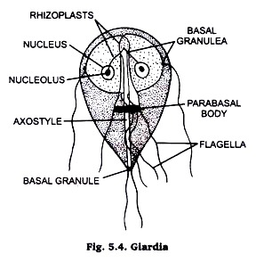
1. The body is bilaterally symmetrical and pear-shaped in appearance with the anterior end rounded and posterior end tapering.
2. The dorsal surface is convex and the ventral surface is flattened provided with a concave sucker.
3. Axostyles form the median longitudinal axis of the body.
4. Four pairs offiagella rise from four pairs of basal granules.
5. Protoplasm contains two nuclei, two axostyles and two parabasal bodies.
6. Reproduction takes place by longitudinal binary fission and cyst formation.
7. The disease caused by this parasite is called giardiasis.
8. Its life cycle is monogenetic and infection to man occurs by the ingestion of its cysts with contaminated food and water.
Habit and habitat:
Giardia is found a parasite in the digestive tract of vertebrates. G. intestinalis is a parasite in the intestine of man.


