In this article we will discuss about the dissection of Wallage Attu.
Procedure for dissection of Wallage Attu:
Take either a freshly-killed or preserved fish and wash it thoroughly if the fish is preserved. Make a longitudinal incision in the integument at mid-dorsal region of the head from pectoral fin to snout.
Likewise, make a transverse incision in the integument at the level of pectoral fin to the mid-ventral line of the lower jaw. Remove the integument so that the half dorsal, lateral and ventral parts of the head are fully exposed.
ADVERTISEMENTS:
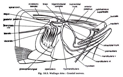
Now, break off the cranium carefully to expose the brain. Then, trace the cranial nerves from their origin to their innervation. When your dissection is complete, display the nerves by placing black paper below them. Draw a neat and well-labelled diagram of the same in your practical record with the help of Fig. 18.5.
There are 10 cranial nerves on one side, thus, there would be 10 pairs of cranial nerves, which arise from the brain and symmetrically disposed on the two sides of the head. The students are usually asked to expose V, VII, IX and X cranial nerves.
The various cranial nerves are as follows:
ADVERTISEMENTS:
I. Olfactory:
Arises from olfactory lobe and innervates the olfactory folds.
II. Optic:
Arises from the optic thalamus of diencephalon, enters the eye ball and supplies to retina.
ADVERTISEMENTS:
III. Oculomotor:
Arises from the ventral aspect of midbrain and supplies to eye muscles.
IV. Trochlear:
Arises from the dorso-lateral aspect of the optic lobe and cerebellum, and innervates the eye muscles.
V. Abducent:
Arises from the ventral side of medulla and innervates to eye muscle.
VI. Trigeminal and VII. Facial:
Arise from the side of medulla, join immediately to form a trigeminofacial complex. The complex separates intracranial into three braches. These are:
1. Supraorbital trunk:
ADVERTISEMENTS:
Which runs forward and forms inner ophthalmicussuperfacialis and outer ophthalmicussuperficialis. These supply to the skin of snout.
2. Infra-orbital-trunk:
Which runs forward and outward and divides into maxillaris, buccalis and mandibularis. Maxillaris supplies to maxillary barbel, buccalis supplies to the skin of snout and also to maxillary barbel, while mandibularis bifurcates to give rise mandibularisexternus supplying to overlying skin of the anterior end of madible and mandibularisinternus supplying to the mandibular barbel and anterior end of head.
The infra-orbital-trunk, before dividing into above 3 branches, also give off following two branches:
(a) pre-maxillaris supplying to the upper jaw.
(b) palatinus anterior which runs on the roof of buccal cavity.
3. Hyomandibular-trunk:
Which immediately gives out a stout opercula is nerve supplying to operculum and then divides into hyoidean and mandibularis branches. The hyoidean branch supplies to the branchiostegal membrane on under-side of the head.
The mandibularis supplies to the lower lip and mandibular teeth. Hyomandibular trunk immediately after its origin from the complex also gives off a slender palatinus posterior nerve which also supplies to the roof of the buccal cavity. From trigeminofacial complex also arises a lateralis accessory nerve which receives dorsal rami of spinal nerves.
VIII. Auditory:
Arises from the lateral side of medulla and divides into vestibular branch supplying to the utriculus and saccular branch supplying the sacculus, lagena and sinus endolymphaticus.
IX. Glossopharyngeal:
Arises from the ventro-lateral side of medulla behind the origin of auditory nerve. It has only post-trematic-branch, enters the first gill and gives a branch to the roof of pharynx.
(i) Branchialis, these are 4 in number. Each with pre- and post-trematic branches supplying to the gills.
(ii) Visceralis, arises from behind the fourth branchialis, which gives a branch supplying to the pericardium and heart, and other branch to the viscera.
The lateralis nerve originates from close to the origin of vagus nerve from medulla and runs back to the posterior and of tail beneath the lateral line system to which it innervates.
Weberian Ossciles of Wallago:
Procedure for dissection:
Take either a preserved or freshly-killed fish remove the skin and muscles of the posterior region of the operculum. Trace out the air bladder and locate a triangular piece of bone attached to its anterior end. This tranangular bone is tripus, a part of weberianossicles. Go on tracing it anteriorly and trace all the related ossicles. Take them out together and place in a watch glass containing water.
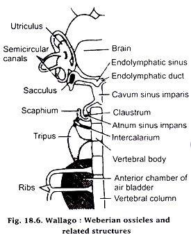
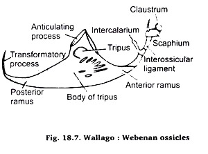
Study it and draw a well-labelled diagram of the same with the help of Fig. 18.6 and 18.7. Weberianossicles are made of a chain of bony structures and are characteristic of order Cypriniforms. These are four bony pieces, claustrum, scaphium, intercalarium and tripus together referred to as Weberianosicles (Fig. 18.7).
The claustrum is the smallest enterior most piece which articulates with neutral arch of first vertebra. The scaphium is a broad and compressed bony piece which is followed by intercalarium and tripus.
Tripus is the largest bony ossicle having three processes, the anterior process is connected to interossicular ligament, medium process is attached to the junction of 2nd and 3rd vertebrae. The posterior process is curved and is connected with the anterior chamber wall of the air or swim bladder. Sound waves are said to travel to the internal ear through these ossicles.
Internal Ear of Wallago: Webebab ossicles:
Procedure for dissection:
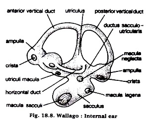
Take a presented or freshly-killed fish and remove its spin from the hinder end of the cranium and exposed its auditory capsule that lodges the internal ear. Carefully scrap the sides of cranium to locate triangular area having three ridges which lodge three semicircular canals.
Now carefully exposed the semicircular canal with the help of scalpel and forceps and also the other parts of the ear. When ear is completely exposed try with a forceps whether it is completely free from the auditory capsule or not? it not then further clear the different parts of the ear.
When it becomes free from auditory capsule take it out and put it in water in a watch glass.


