The below mentioned article provides a study note on Paramoecium.
Introduction to PARAMOECIUM:
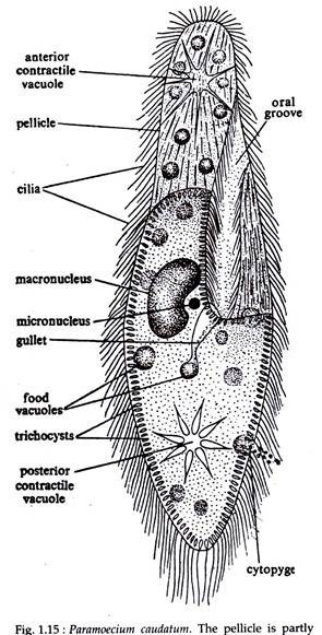
The Phylum Ciliophora, to which Paramoecium belongs, comprises of organisms that is highly differentiated in structure amongst the unicellular Protozoa. They have characteristic hair-like organelles, the cilia, for locomotion and food capture (Fig. 1.15).
In contrast to other protozoans, they have two types of nuclei – one vegetative (the macronucleus), and the other reproductive (the micronucleus). Paramoecium, the name given by John Hill (1932), has a number of species of which P. caudatum is the longest and have a single micronucleus and commonly found P.
ADVERTISEMENTS:
Aurelia is less pointed at the rear end and has two micronuclei, while P. multimicronucleatum has many micronuclei. P. bursaria is easily recognised by its green colour due to the presence of symbiotic algae, Zoo chlorella.
Etymology of PARAMOECIUM:
Greek: paramekes, ablong.
The species name caudatum is derived from the Latin word caudata meaning tail. Paramoecium, due to its shape is sometimes called as ‘slipper animalcule’ or ‘Ladies slipper’.
ADVERTISEMENTS:
Habit and Habitat of PARAMOECIUM:
Paramoecium is found in fresh-water ponds, pools, ditches/streams, rivers, lakes and reservoirs. They are specially abundant in organic infusions, that is, in stagnant water containing decaying organic matter, feeding upon bacteria and tiny protozoa that swarm in such waters.
Structure of PARAMOECIUM:
External Structure:
ADVERTISEMENTS:
Paramoecium has a permanent body shape (Fig. 1.15). Its body is slipper or cigar- shaped and measures about 0-25 mm in length. One end of the body is slender but rounded or blunt, representing the heel of the slipper, is the anterior end.
The other end is thick and pointed, representing the top of the slipper, is the posterior end. The flattened surface of the body which is with a groove that always faces the substratum is called the oral or ventral surface, while the opposite and somewhat convex side is the aboral or dorsal surface.
The body of Paramoecium is covered with a distinct, tough and flexible pellicle. The pellicle is sculptured into hexagonal depressions, provided with holes at the centre through which the cilia projects. In Paramoecium the ciliature is divided into body ciliature, which occur over the general body surface and the oral ciliature which is associated with the mouth region or oral groove and it includes larger cilia.
Internal Structure:
Beneath the pellicle the protoplasm is differentiated into cortex and medulla.
Cortex:
The cortex or ectoplasm is clear, non-granular, less extensive and contains many spindle-shaped bodies called trichocysts. Trichocysts measure about 1 /100 mm in size and are arranged as rows of sacs having their closed sides towards the inner layer.
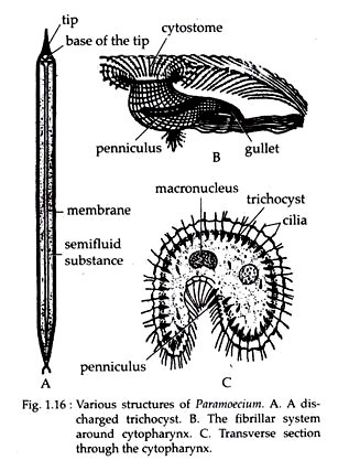
The trichocysts are filled with homogeneous, refractive and semi-fluid substances. On being stimulated the contents come out through small openings on the ridges of the pellicle. When the stimulations are very strong the trichocysts are totally expelled out of the body as long threads (Fig. 1.16A).
ADVERTISEMENTS:
Meulla:
The medulla or endoplasm is granular and bears at the centre the nuclear apparatus, consisting of a large kidney shaped macro-nucleus and a small rounded micronucleus. The micronucleus is placed in the concavity of the macronucleus. The macronucleus and micronucleus are concerned with ordinary life processes and reproductive processes, respectively.
The medulla also contains two star-shaped contractile vacuoles, one situated at the anterior and the other at the posterior end of the body. Many food vacuoles, containing food particles at different stages of digestion, are scattered in the medulla. Posterior to the mouth there is a definite anal spot (cytopyge), through which undigested food particles are eliminated to the outside.
The oral groove, situated on the ventral surface of the body, runs obliquely backward and opens into a funnel-shaped depression called the gullet or cytopharynx (Fig. 1.16B) through an aperture called cytostome. The cilia, lining the gullet are larger in size. A special ciliary structure called penniculus (Fig. 1.16C) is formed by the fusion of four rows of cilia. Two such penniculi are seen in the gullet region.
Locomotion:
Paramoecium exhibits two types of movement—creeping and swimming.
Nutrition:
Paramoecium is typically a holozoic animal. It is a microphagous animal, feeding on small organisms like protozoa, bacteria and minute particles of animal and vegetable matter.
Respiration:
By the process of diffusion, oxygen dissolved in water enters through the surface of the body and carbon dioxide passes out in the same manner.
Osmoregulation and Excretion:
The two contractile vacuoles present at either end of the body serve as effective osmoregulatory organelles in Paramoecium. The detailed structure of the contractile vacuoles and its role in osmoregulation and excretion are described elsewhere.
Reproduction:
Reproduction in Paramoecium takes place by both asexual and sexual means.
Irritability:
The behaviour of Paramoecium to various kinds of stimuli are interesting.
Some of the responses are dealt below:
i. Response to Mechanical Stimuli:
The anterior end of Paramoecium is more sensitive than the posterior end and offers either positive or negative response. The response to contact or thigmotropism is, therefore, varied.
ii. Response to Gravity:
Paramoecium exhibits negative reaction to gravity or geotropism and in a culture jar, resides just below the surface of the medium.
iii. Response to Temperature:
Response to temperature is known as thermo-tropism. Paramoecium enjoys an optimum temperature between 24°C and 28°C. It avoids any rise or lowering of this temperature.
iv. Response to Light:
Response to light is known as phototropism. Paramoecium exhibits negative response to strong light and ultra violet rays.
v. Response to Chemical Stimuli:
Paramoecium exhibits negative chemotropism or reaction to chemicals. It thrives in a drop of medium containing 0-02 per cent acetic acid but if the concentration of the acid is increased the animal moves away to a place where the acid is diluted by the surrounding water.
vi. Response to Water Current:
Positive Theo-tropism is shown by Paramoecium. It orients itself with its anterior end upstream while swimming against the current.
vii. Response to Electrical Stimuli:
Positive Galvan tropism is exhibited by Paramoecium to a weak electric current with the animal moving towards the cathode. During strong electric current, the animal, however, moves backward towards the anode, where it finally disintegrates and dies.
The above described sensitivity and movements of Paramoecium prove that a certain level of sensory organelles has developed in it. Recently acetyl cholinesterase activity has been reported from the base of each cilia which is a neurohormone (acetylcholine) breaking enzyme.

