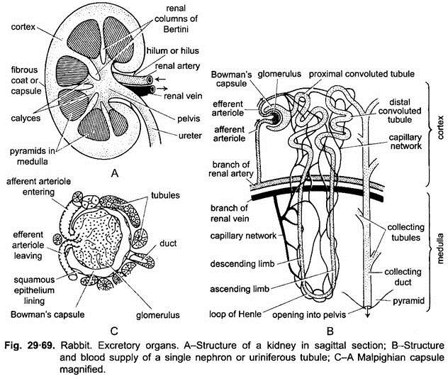
This system is generally responsible for the elimination of metabolic waste products (nitrogenous waste products in the form of urea, etc.), excess of salts and water from the body. The organs of excretion in rabbit are a pair of kidneys, ureters and a urinary bladder.
Kidneys:
The kidneys are metanephric and main organs of excretion.
1. External Structure:
The kidneys are two, dark red and bean-shaped attached to the dorsal abdominal wall outside the coelom, one on either side of the vertebral column. The right kidney is situated somewhat more anterior than the left. Each kidney is ventrally covered by peritoneum. The outer side of each kidney is convex and inner side is concave having a notch or depression, the hilum or hilus. A renal artery enters the kidney at hilus and a renal vein comes out of it through the hilus.
ADVERTISEMENTS:
2. Internal Structure:
In a longitudinal section, the mammalian kidney is seen enclosed in a thin capsule of fibrous connective tissue.
Cortex:
Beneath the capsule is the cortex part of the kidney which is homogeneous and light in colour.
ADVERTISEMENTS:
Medulla:
This is the inner region of kidney which is dark in colour.
Pelvis:
It is a large, funnel-shaped space in the centre of kidney towards the hilus. The urine is collected here and later drained off by the ureter.
ADVERTISEMENTS:
Pyramids:
In rabbit and man, the medulla is formed of a number of lobes, called pyramids, and the cortex is continued inside between the pyramids to form renal columns of Bertini. In rat the conical pyramid is single. The pyramids are projected into the pelvis. The kidney is formed of a mass of fine, long, convoluted tubules, called the uriniferous tubules or nephrons embedded in connective tissue having blood and lymph vessels, nerves and smooth muscle fibres.
Each uriniferous tubule is formed of a Malpighian capsule and convoluted duct.
The Malpighian capsule consists of a proximal cup-shaped Bowman’s capsule in the cortex and in its lumen is present a tuft of blood capillaries, called glomerulus. The Bowman’s capsule enclosing the glomerulus is collectively known as Malpighian capsule or renal corpuscle.
The convoluted tubule behind the Malpighian capsule is divisible into three regions:
(a) Proximal convoluted tubule,
(b) Henle’s loop and
(c) Distal convoluted tubule.
ADVERTISEMENTS:
The proximal convoluted tubule starts from the Bowman’s capsule and makes a few coils in the cortex and then proceeds downwards as descending limb in the medulla to form a thin loop of Henle. It again proceeds towards the cortex as ascending limb and again extends towards the cortex as ascending limb and forms a few coils in the cortex, called the distal convoluted tubule.
The distal convoluted tubule joins with one of the larger collecting ducts. The collecting ducts collectively form a pyramid and finally open into the pelvis. The wall of the tubule is made of cuboidal epithelial cells which are ciliated at interval, whereas the wall of Bowman’s capsule is formed of single layer of squamous epithelium.
Blood supply of the kidney- The renal artery after entering into the kidney divides and redivides forming several arterioles. Each arteriole enters a Bowman’s capsule as an afferent arteriole which capillarises to form the glomerulus and then an efferent arteriole emerges from the glomerulus and again capillarises around the convoluted tubule to distribute the blood in the remaining part of the tubule.
These capillaries unite together to form a venule and a number of such venules unite together to form a renal vein which comes out of the kidney from the hilus. The diameter of efferent arteriole is larger than the efferent arteriole. Branches of renal artery and renal vein run along the junction of cortex and medulla and give branches to the glomerulus and receive branches from them respectively.
Ureters:
Each thick-walled ureter starts from the hilus and runs backwards along the dorsal abdominal wall and opens posteriorly into the neck of urinary bladder.
Urinary Bladder:
The urinary bladder is a pear-shaped, transparent muscular sac, situated in the pelvic region ventral to the rectum and connected to the ventral abdominal wall by a suspensory ligament. It serves as urine reservoir.
The posterior narrow neck of the bladder bears a circular sphincter muscle. The neck of urinary bladder opens into a thick-walled, muscular canal, the urethra. In male, it is much longer and passes through the penis and opens at its tip. It is called urinogenital canal. In female, it is short and unites with the vagina to form the vestibule and opens out by a slit-shaped vulva.
Physiology of Excretion:
The nitrogenous waste products of metabolism in rabbit are generally urea which is synthesised in the liver from amino acids, by a cyclical chain of reactions. The urea is then carried through the blood into the kidneys where it is eliminated with the urine. Urine formation in kidneys occurs by ultrafiltration, selective reabsorption and secretion.
1. Ultrafiltration in Glomerulus:
Blood comes under high pressure in the glomerulus through the afferent renal arteriole whose diameter is greater than that of efferent arteriole. Here ultrafiltration occurs through the permeable walls of the capillaries of glomerulus.
Thus, due to ultrafiltration the dissolved substances of the blood, e.g., urea, salts (sodium and potassium), glucose, water, etc., are filtered into the lumen of the cup of Bowman’s capsule. Now the blood contains corpuscles, proteins and fats. This filtrate is known as glomerular or capsular filtrate. The remaining blood constituents pass into the efferent arterioles. This process is simply a physical process.
2. Selective or Tubular Reabsorption:
The glomerular filtrate contains some useful substances like water, glucose, amino acids and some salts. These useful substances are reabsorbed in the proximal convoluted tubules from the glomerulur filtrate. The absorbed substances then pass into the blood circulation through capillaries of the efferent arterioles found around the convoluted tubule which finally joins to form renal venules.
The maximum water is reabsorbed in the loop of Henle and no reabsorption occurs in the distal convoluted tubule. Glucose, amino acids and some urea are reabsorbed in the proximal convoluted tubule, while chloride and bicarbonate of sodium are reabsorbed in the proximal and distal convoluted tubules. The water and urea are reabsorbed by passive diffusion. Amino acids, salts and sugars, etc., are reabsorbed by active transport.
Thus, as the glomerular filtrate passes through the convoluted tubule, the useful substances are reabsorbed and the concentration of waste products including urea increases.
3. Tubular Secretion:
In this process some substances like remnant urea, creatine, creatinine, ammonia, hydrogen, potassium ions, various drugs, etc., are secreted into the lumen of tubule from the efferent capillaries surrounding the tubules.
After tubular secretion the substance which is left in the tubule is rich in excretory wastes and finally known as urine which contains nearly 95% water, 2% urea, 6% Ca and Na chlorides, some uric acid, creatinine, ammonia, etc. It also contains a pigment, urochrome, formed by the breakdown of haemoglobin. It gives yellow colour to the urine.
The urine, thus, formed passes into the collecting tubule and finally reaches the pelvis and ureter from where it is collected in the urinary bladder. The bladder contracts and forces the urine outside through the urinogenital canal and urinogenital aperture.
Besides excretion, kidney regulates the body fluids (blood and water) within the body, thus, also acting as homeostatic organ. Homeostasis is the method for regulating concentrations and contents within the body to preserve equilibrium in any animal.
4. Other Organs of Excretion:
Besides kidney, some other organs also perform excretory functions. The CO2 evolved during tissue respiration is removed by the lungs. Some amount of water is removed in the form of vapours with the expired air and some salts are also removed with the sweat. Bile salts and bile pigments are removed by the alimentary canal which are produced in the liver.
