The sexes are separate and sexual dimorphism is well marked in rabbit.
Male Reproductive System:
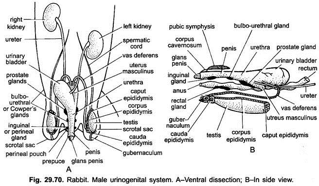
Male reproductive system (Fig. 29.70) consists of a pair of testes, a pair of vasa deferentia, uterus mascuiinus or seminal vesicle, urethra, penis and a number of accessory glands like prostate, Cowpers, perineal and rectal glands.
1. Testes:
ADVERTISEMENTS:
The testes are paired, light pink-coloured, ovoid bodies. These lie in the foetus in the abdominal cavity near the kidneys. But at puberty these descend outside from the body cavity through inguinal canals into a special part of the coelom, the scrotal sacs.
The scrotal sacs are covered externally by a thin hairy skin, situated ventrally in the pubic region on either side of the penis. In most mammals, testes remain within scrotal sacs throughout life. But in rabbit, rat and other rodents, they remain within scrotum only during breeding season.
In non-breeding season they are withdrawn into the abdominal cavity through the inguinal canals, thus, these canals remain open throughout life in these animals. It is due to temperature difference, abdominal cavity temperature is higher than that of scrotal sacs.
Development of sperms needs a slight lower temperature, so they descend in scrotum during breeding season. The cavity of the scrotal sacs is connected with the abdominal cavity by a narrow passage, known as inguinal canal.
ADVERTISEMENTS:
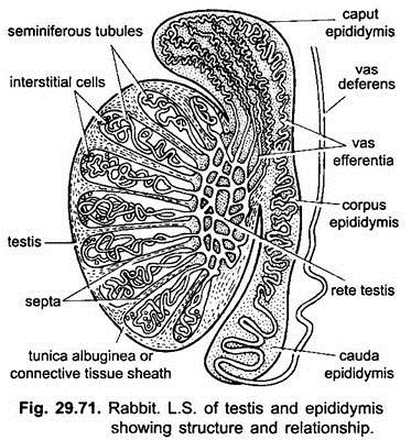
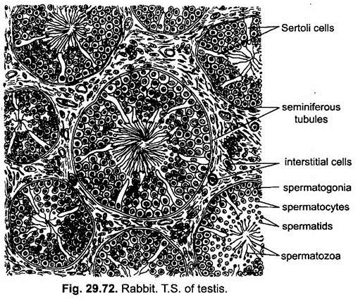
Histologically, each testis is covered by a tough coat of fibrous connective tissue called tunica albuginea, which is projected into the substance of testis in the form of a number of septa forming a number of lobules within the kidney. Each lobule is occupied by long convoluted seminiferous tubules which are bound together by connective tissue.
The seminiferous tubules are lined by germinal epithelial cells which are of two types- smaller, more numerous are the spermatogonial cells which produce spermatozoa after spermatogenesis, and others are a few large-sized columnar supporting cells, called Sertoli cells. These nourish the spermatozoa before they leave the tubule.
The connective tissue having interstitial cells or cells of Leyding are found in between the seminiferous tubules. These cells are endocrine in function and secrete hormones, which control the secondary sexual characters.
In each testis, all the seminiferous tubules open into a network, called the rete testis. It communicates by many fine ductules, the vasa efferentia into the epididymis. The vasa efferentia are lined internally by ciliated epithelium.
ADVERTISEMENTS:
2. Epididymis:
It is a long, narrow and highly convoluted tubule forming a compact protrusion all along the inner surface of the testis. The epididymis is divisible into three regions anterior caput epididymis or globus major, middle narrow corpus epididymis and posterior cauda epididymis or globus minor.
Caput Epididymis:
It is the anterior part of epididymis and connected with the testis by a number of vasa efferentia. It is also connected with the dorsal abdominal wall by a spermatic cord formed of connective tissue, artery, a plexus of vein around the artery, called pampiniform plexus. The cauda epididymis is connected with the scrotal sac by a thick connective tissue cord known as gubernaculum.
Corpus Epididymis:
It is the middle narrow part which connects the anterior and posterior part of epididymis. Caput and cauda epididymes store and nourish the sperms.
3. Vasa Deferentia:
From the cauda epididymis of each testis arises a straight, muscular tube, the vas deferens or sperm duct. It passes forwards along the inner side of the scrotal sacs and then enters the abdominal cavity through the inguinal canal.
ADVERTISEMENTS:
Then it curves over the ureter and passes backwards to open in a small median bag, called uterus masculinus or seminal vesicle situated above the neck of the bladder. The seminal vesicle opens dorsally into urethra. Urethra is the distal narrow part of the urinary bladder. The urethra, thus, serves as a passage for urine and the spermatic fluid. It opens at the tip of penis.
4. Urethra:
The neck of urinary bladder and the seminal vesicle open into a thick- walled muscular duct, called the urethra or urinogenital canal.
5. Penis:
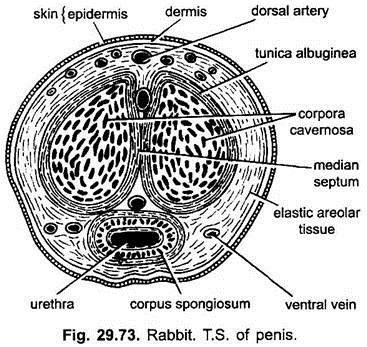
The penis is a muscular, cylindrical, and vascular erectile organ, which hangs in front of the anus. The penis is covered by a loose sheath of skin which hangs freely over and beyond the tip of the penis as a prepuce. The cap-like tip of the penis covered by prepuce is called glans penis.
The penis consists of three spongy, vascular longitudinal columns around urethra, called corpora spongiosum and above it two corpora cavernosa. These spongy muscles are surrounded individually by tunica albuginea, a fibrous connective tissue layer. Around these three columns is present elastic areolar tissue layer containing blood vessels and nerves. The blood spaces of the penis get filled with the blood at the time of sexual excitement to make the penis stiff and enlarged.
6. Accessory Glands:
A number of accessory sex glands open into the urethra. Their secretions, along with those of epididymis, seminal vesicles form the seminal fluid (semen).
These sex glands are as follows:
(i) Prostate Gland:
A large prostate gland is situated dorsally at the base of uterus masculinus which opens into the urethra by numerous ducts. Its secretion is white and alkaline, which makes the passive sperm active.
(ii) Cowper’s Glands or Bulbo-Urethral Glands:
A pair of Cowper’s glands is situated just anterior to the prostate gland on urethra. Their secretion neutralises the acidity and, thus, protects the sperms.
(iii) Perineal Glands:
A pair of elongated scent glands lie anterior to the Cowper’s glands. They open into the hairless perineal depressions one on either side of anus. Their secretion gives a characteristic odour of rabbit.
(iv) Rectal Glands:
A pair of rectal glands lie on the dorsal side of rectum. Their function is not known. These two last glands are not associated with the male reproductive organs.
The secretions of prostate and Cowper’s glands activate and nourish the sperms, provide a fluid media for the flow of sperms and neutralise the acidity of the urethral passage.
Female Reproductive System:
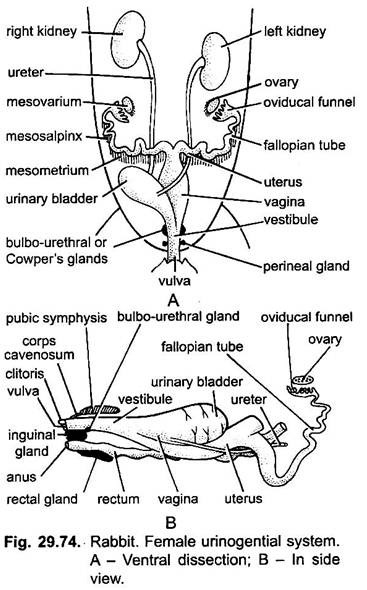
Female reproductive system (Fig. 29.74) consists of a pair of ovaries, a pair of oviducts, uterus, vagina, vestibule and some accessory glands.
1. Ovaries:
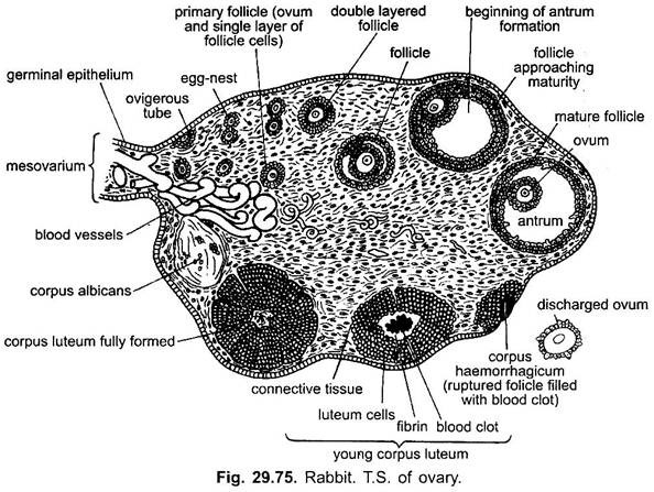
The paired ovaries are small, whitish, oval bodies found attached to the dorsal wall of the abdomen behind the kidneys. The ovaries are attached with the dorsal body wall by a double fold of mesentries, called mesovarium. Each ovary when ripe shows on its outer surface many tiny projections called ovarian or Graafian follicles. Each follicle contains a developing ovum. These projections rupture releasing the ova.
Histological, each ovary is made up of a dense fibrous connective tissue called stroma. It contains blood and lymph vessels, nerves and follicles in various stages of development. The stroma is surrounded by a layer of germinal epithelial cells and internal to it is present a thin layer of connective tissue, called the tunica albuginea.
Stroma is further differentiated into outer broad cortex and inner narrow medulla containing only blood and lymph vessels. The cortex contains ovarian follicles in various stages of development and regressing follicles like corpora lutea and corpora albicans and interstitial cells. One follicular cell develops to become an ovum and the other cells called discus proligerous around it protect and nourish the ovum. It is attached to one side of the follicle.
In a mature Graafian follicle, a fluid-filled cavity appears which separates an outer layer of cells, the membrana granulosa, from the cells of discus proligerous. The follicular cells around the ovum secrete a thick membrane called zona pellucida. It is covered by another membrane of striated columnar cells, called corona radiata. Such a mature follicle is known as a Graafian follicle.
The Graafian follicle migrates towards the surface of the ovary and ruptures to release the ovum (ovulation). After the release of the ovum is left a mass of follicular cells surrounding a blood clot. Gradually these follicle cells proliferate rapidly and transform into a yellow coloured mass of cells, the corpus lutem, which produces a hormone, progesterone.
It is responsible for preparatory changes in the uterus for fertilisation and retention of the embryo during gestation. Thus, the corpus luteum remains active throughout the period of embryonic development. If, somehow, no fertilisation occurs, it degenerates leaving a scar, called corpus albicans.
2. Oviducts:
There is a fimbriated (lined by cilia) funnel or ostium close to the outer side of each ovary which leads into the oviduct. The fimbriated funnel receives the ovum after their discharge from the ovary. Each oviduct behind the funnel is differentiated into two parts- first part, the follopian tube, is coiled, narrow, internally ciliated and glandular and the second part is long, thick, muscular, glandular and coiled with wider diameter, called uterus.
Both the uteri are attached with the dorsal abdominal wall by a mesentery. Fertilised ova are implanted on the uterine wall for the development. The developing embryo remains attached with the uterine wall by a placenta.
3. Vagina and Vestibule:
The uteri of both the sides join together along the median line to form a common wide passage, called the vagina. It lies over the urinary bladder. The vagina passes backwards and joins with the neck of the urinary bladder, forming a common passage, called urinogenital canal or vestibule. It extends backwards ventral to rectum and opens outside by a slit-like aperture, the vulva.
4. Clitoris:
It is small, erectile, and knob-like, and attached to the anterior wall of vulva. It is homologous to the penis of male because it consists of a pair of erectile tissue (corpora cavernosa) which is highly sensitive.
5. Accessory Glands:
A pair of small Bartholin’s glands is found embedded in the dorsal wall of vestibule. The secretion of these glands is discharged in the vestibule which lubricates the vaginal passage. They are similar to the Cowper’s glands of the males. The prostate glands are not found in female rabbit. A pair of perineal glands and rectal glands is also found like those of the male rabbit.
Copulation:
The domestic rabbits can copulate at any time but wild rabbits copulate from January to June. Usually a female in most mammals copulates only when she is in oestrus condition or on heat, of which rabbit may be an exception. At the time of copulation the penis of the male is introduced in the vagina of the female where semen is ejaculated.
Fertilisation and Development:
After about 8-10 hours of copulation, the Graafian follicles mature and ovulation occurs. The ova discharged from the Graafian follicle are immediately caught by the ostium and then passes down the fallopian tubule where fertilisation occurs. The ova undergo first maturation division before their discharge from the ovary. The second maturation division occurs at the time of fertilisation after the entry of sperm in the ovum.
At the time of fertilisation the entire sperm enters the ovum but its tail degenerates. The penetration of sperm into ovum is helped by an enzyme hyaluronidase secreted by the acrosome of the sperm. Usually only one sperm enters the ovum. After the entry of sperm in the ovum, the head of sperm rotates through 180 degrees and finally the male and female pronuclei fuse together resulting into the formation of zygote nucleus.
The zygote then passes down the fallopian tube and reaches the uterus where it gets attached to the wall of the uterus. After next few days a special connection placenta is formed between the developing embryo and uterine wall. The placenta helps the embryo in all the physiological exchange of materials with the mother until birth. The gestation period in rabbit is about 28-32 days, after which mother gives birth to the young ones.
