The sense organs or receptor organs of rabbit are essentially similar with that of the frog except in some details. The sense organs detect the changes in the external and internal environments. The changes in the environment are known as stimuli.
The various sense organs of rabbit are:
1. Organs of touch — Cutaneous (Skin) receptor.
2. Organs of taste — Gustatoreceptors (Tongue)
ADVERTISEMENTS:
3. Organs of smell — Olfactoreceptors (Olfactory sacs or nasal chamber)
4. Organs of sight — Photoreceptors (Eyes)
5. Organs of hearing and equilibrium — Statoacoustic receptors (Ears)
1. Organs of Touch (Skin):
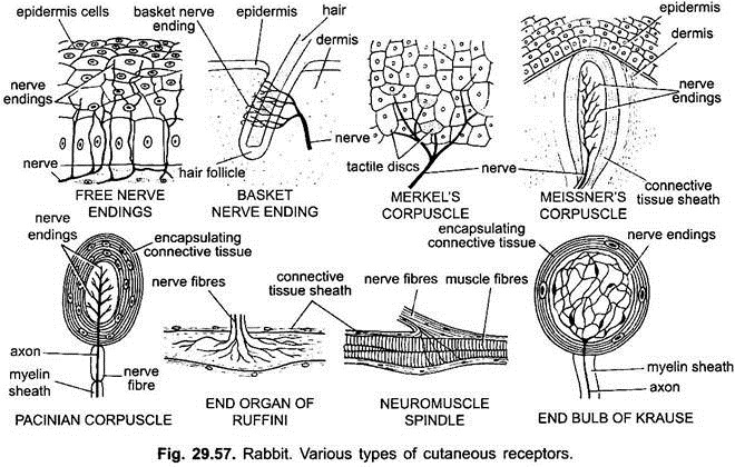
The skin of mammals is highly sensitive which is provided with several types of receptors. These receptors are microscopic and are of various types situated beneath the epidermis. Each receptor is concerned with a particular stimulus.
ADVERTISEMENTS:
i. Free Nerve Endings:
Sense of touch, cold, warmth, pressure and pain, etc., are perceived by free nerve endings. The fine branching fibres of sensory nerves lie just beneath the epidermis of skin. These fibres also extend into the epidermis.
ii. Basket Nerve Endings:
These are concerned with sense of touch. A fine network of branching sensory nerve fibres are found around the hair follicle. Sense of touch is perceived by touching the hair.
ADVERTISEMENTS:
iii. Encapsulated Nerve Endings:
These are in the form of capsules, e.g., Meissner’s and Pacinian capsules. Meissner’s capsules are found just beneath the epidermis. There are formed of a naked axon surrounded by a sheath of connective tissue capsule. These are sensitive to touch. Pacinian capsules are found in dermis and several internal organs. Each capsule is formed of a core of single axon ending in an ovoid bulb, which is surrounded by a sheath of connective tissue. These detect pressure.
iv. Neuromuscular Bundles or Spindles:
These are found inside the body, i.e., in skeletal muscles, tendons and joints. These are called kinaesthetic receptors and apprise the central nervous system about the degree of muscle tension. It is formed of nerve ending in bulb or end organ, called the neuromuscular bundle or neuromuscular spindle.
The tactile organs are characteristically known as corpuscles. These corpuscles are ovoid bodies supplied with nerve-endings. Each corpuscle is formed of a sheath of connective tissue arranged in concentric rings around the core of a nerve fibre.
The corpuscles are tangoreceptors, frigidoreceptors, caloreceptors and algesireceptors, i.e., sensitive to touch, cold, warmth or heat and pain respectively. However, the pain receptors (algesireceptors) are without special corpuscles and only in the form of fine nerve fibres. These corpuscles are also responsible for sensitiveness to humidity and pressure, etc.
2. Organs of Taste (Gustatoreceptors):
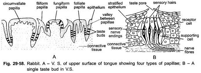
These are the taste-buds found on the tongue papilla and on the soft palate (roof of the buccal cavity). The taste bud is barrel-shaped and made of two kinds of cells, the neurosensory cells (gustatory cells) and supporting cells. The gustatory cells are elongated, spindle-shaped and provided with sensory hairs projecting in a depression between tongue-papillae called taste-pore.
The inner ends of neurosensory cells are supplied by the nerve fibres from VII and IX cranial nerves. These cells are stimulated by the substances dissolved in the mucous and saliva and the sensation of bitter, sweet, salty, sour, etc., finally reaches to the brain.
3. Organs of Smell (Olfactoreceptors):
ADVERTISEMENTS:
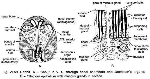
The nasal passage is provided with scroll-like turbinal bones, known as ethmoturbinals, maxilloturbinals and nasoturbinals on the basis of their origin. These turbinals and roof of nasal chambers are covered over with olfactory epithelium or Schneiderian epithelium. The olfactory organs have spindle-shaped olfactory cells, mucous cells and columnar supporting cells.
The olfactory cells externally bear several delicate olfactory hairs and their inner ends are connected with nerve fibres which enter the olfactory lobes of the brain. The mucous cells produce mucus which keeps back particles of dust and it also dissolves odoriferous substances which are in gas form and stimulate the neurosensory cells or olfactory cells. The sense of smell is well developed in rabbits.
The rabbit in embryonic condition bears Jacobson’s organ in the roof of buccal cavity, in the form of a groove on the lower medial aspect of each nasal cavity. Its duct is separate from the nasal apparatus and opens into the mouth cavity. It is not found in adults.
The epithelium of ethmoturbinals actually constitutes the olfactory region which is sensory to smell. The epithelium of maxilloturbinal moistens and warms the air on its way to lungs and is not sensory. The epithelium of nasoturbinals has no olfactory cells.
Each nasal passage has an ovoid sensory pad having minute papillae and ridges. These pads are tactile and serve as distance receptors for testing air.
4. Organs of Sight or Eyes:
The eyeballs are spherical photoreceptor organs, situated one on either side of the head in the orbits. The eyes of mammals are similar in their structures.
External Structure:
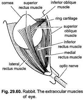
The eyeball is spherical and hollow. Its about one-fifth part is visible externally and the remaining part remains hidden within the body orbit. Each eyeball is provided with six sets of muscles which move the eyeball in the orbit.
Out of the six sets, four sets are rectus muscles and two sets are oblique muscles. These muscles are attached on one side with the eyeball and on the other side with the orbit. The exposed part of the eyeball is known as cornea which is well protected by two movable eyelids the upper and lower. Both the eyelids are provided with stiff hairs or eyelashes on their margins.
These eyelashes cornea protect the eyes from dust particles, rain water and sweat, etc. A transparent nictitating membrane, the third eyelid is present in inner corner of the eye and can cover the whole cornea in rabbit. Its function is to clean and protect the eye from dust particles. In human beings it is rudimentary in the form of pink mass.
Glands:
There are three types of glands in each eye- Meibomian, Harderian and lacrimal.
Meibomian glands are sebaceous glands placed in both the eyelids beneath the conjunctiva and open on the free edges of eyelids. They secrete an oily secretion for lubricating the eyelids. The Harderian glands are situated at the inner side below the lower eyelids and open in connection with nictitating membrane at the inner angle, whose secretion keeps the conjunctiva moist.
The lacrimal gland has several openings on the conjunctival surface below the upper eyelid, towards the outer side of eye. These glands secrete a saline watery fluid, the tears, on the surface of the eye (conjunctiva). It keeps the eye moist, soft, clean and free from bacteria. The excess of tears accumulate towards the inner comer of eye and are drained by a naso-lacrimal duct into the nasal chamber. Harderian glands are absent in primates.
Internal Structure:
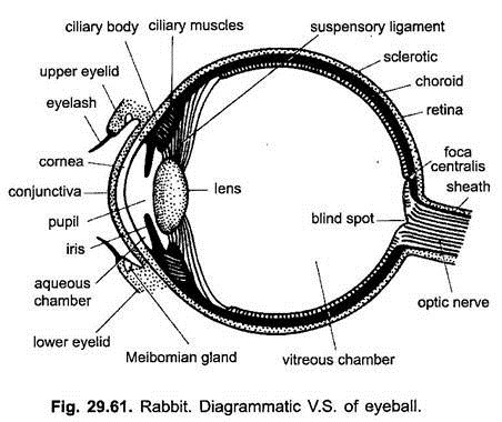
The internal structures of the eyeball can be well explained with the help of its vertical section as shown in the diagram. Its wall consists of three coats- an outer sclerotic, middle choroid and inner retina.
(i) Sclerotic:
The sclerotic is a tough layer of thick, white and opaque dense fibrous connective tissue. The sclerotic layer maintains the form of the eyeball and covers the greater part of it. The front exposed part of sclerotic layer is transparent through which light enters, and bulged a little to form the cornea.
The cornea is covered by a thin delicate, transparent, epithelial layer, the conjunctiva. The conjunctiva is continuous with the epidermis lining the eyelids. It contains blood capillaries and free nerve endings.
(ii) Choroid:
The choroid cornea is the middle layer formed of conjunctiva loose, pigmented and highly vascular connective tissue. This layer helps in darking the cavity of the eyeball to check the internal reflection of the light. Its blood capillaries provide nourishment to the retina.
The choroid is found beneath the posterior part of sclera. In front at the junction of sclerotic and cornea the choroid swells and bends inwards into the cavity of the eyeball forming the ciliary body which is provided with ciliary muscles which project into vascular ciliary processes or folds. These secrete aqueous humour.
In front of ciliary body, the choroid becomes separate from the cornea or at the corneal margin the choroid is continued as iris, which is separated by a space (anterior chamber of eye) from the cornea. In this region its pigmentation gives the characteristic colour to the eye. It is perforated by a circular aperture, the pupil. In the iris are present two sets of smooth muscle fibres- radiating fibres for dilation and circular fibres for contraction of the pupil. Thus, the diameter of the pupil is controlled by the contraction and relaxation of the muscles of the iris.
Lens:
Just behind the iris is a solid biconvex lens enclosed in a lens capsule. Lens is formed of concentrically arranged transparent fibres. Lens capsule is thin, transparent and elastic. Lens is supported by the suspensory ligaments attached with the ciliary bodies.
(iii) Retina:
The retina is the innermost transparent layer of the eyeball whose outer layer is pigmented and closely applied with the choroid and the inner nervous layer which is sensory layer. The nervous layer is provided with an outer layer of rods and cones which are photoreceptor cells. The outer pigmented layer is of simple cuboidal or columnar epithelium. The inner layer is also a simple cuboidal epithelium in the pars iridica (beneath the iris) and in the pars ciliaris (in the region of ciliary body).
Retina beneath the iris is also pigmented. This part of retina is not light sensitive and relatively simple in structure. Only the posterior part of retina (pars optica) is light sensitive and highly complex, containing light-sensitive elements and nerve cells and fibres.
Light- sensitive elements are rods and cones. Their inner ends connect with small bipolar nerve cells which in turn connect with large nerve cells (ganglion cells). From the ganglion cells arise fibres, all of which converge on a small circular area, towards nasal side of the posterior pole of eyeball, called optic disc, and here they pierce the coats of eyeball to become optic nerve.
The rods are more sensitive to low intensity of light and contain a pigment rhodopsin. They are suitable for dark (night vision). The cones are sensitive to high intensity light and more suited for day vision. They produce a sharp image. Cones are also sensitive to light of relatively narrow frequency bands. Thus, they are associated with recognition of colours, which is only found in primates.
Rods are more towards the periphery, while cones are more concentrated towards the centre. The site where all the nerve fibres converge and unite to form the optic nerve and then leave the eye is called the blind spot.
It is without any photoreceptor cells and, therefore, does not produce any impression. Just above the blind spot in the retina in line with the optical axis is a slight, oval, depression called yellow spot or area centralis. Moghe (1957), however, has described that yellow spot is not found in the eyes of rabbit. The oval depression in the area centralis, called fovea, is the region for most distinct vision. Here rods are absent and only cones are present.
Chambers:
The cavity of the eyeball is divided into an anterior and a posterior chamber by the iris, lens and suspensory ligaments. The anterior chamber between lens and cornea is the small aqueous chamber filled with a watery fluid, the aqueous humour. The posterior chamber uses between lens and retina is large, called vitreous chamber.
The vitreous chamber is filled with a gelatinous fluid, the vitreous humour. The aqueous humour is secreted by the ciliary body and carried through canal of Schlemm at the base of cornea. It nourishes cornea and lens and maintains intraocular pressure. The disease glaucoma is resulted due to imbalance of intraocular pressure. It damages the retina.
Working of the Eye:
Image Formation:
The cornea, aqueous humour, lens and vitreous body together constitute the dioptric apparatus, which focuses an image of external objects on the retina. The iris is a diaphragm by which amount of light which enters can be regulated. The eye works exactly like that of a photographic camera.
The light rays reflected from an object are refracted by the cornea and the rays are then focussed by the lens on retina, so that a sharp, inverted image is formed. The inverted retinal image is interpreted by the brain and the real sensation of sight arises and the animal sees the object in an upright way. However, the inverted retinal image is never reinverted by the brain.
The type of vision which is found in higher mammals is called binocular (stereoscopic) vision. In such a vision the fields of vision of the two eyes overlap or even coincide each other and, hence, two images of the same object are not seen. Thus, both the eyes can see a single object in three dimensions, and the animal can estimate the distance also. This type of vision in which distance is also estimated is called binocular vision.
But in the lower mammals like rabbit, both the eyes have divergent axes of vision because the fields of vision do not overlap each other. Therefore, the rabbit can see all around by its two eyes, each eye covers a different field of vision. Such type of vision is called monocular vision.
Accommodation or Focusing:
The power of accommodation is well exhibited by the eyes of rabbit for the objects situated at various distances. In other words, it can be said that the power of accommodation is an adaptation of seeing the objects situated at various distances. The accommodation is brought about by changing the convexity of the lens because the distance between lens and retina is fixed and unchangeable. Thus, change in convexity of lens is essential for the formation of distinct images on the retina.
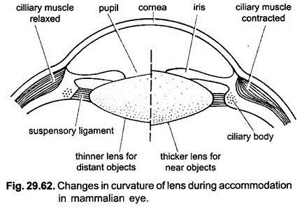
In normal condition when the eyes are at rest, the lens is kept flattened by the suspensory ligaments and, thus, it is adjusted for seeing distant objects. It is due to relaxed condition of the circular muscles of ciliary body. The diameter of the ciliary body is also much increased due to outward pressure of the fluid of eyeball. It, thus, increases the tension of the suspensory ligament, which pulls the lens capsule making the lens thinner or flattened.
When objects from near are to be viewed, then the ciliary muscles contract, thus, reducing the diameter of the ciliary body. Suspensory ligaments also become relaxed due to lessening of tension over them. Thus, the lens becomes thicker and more convex. The cornea also arches outwards. Thus, the focal length of the lens is reduced and a clear image of the near object is formed on the retina.
Chemistry of Vision:
The rods in rabbit (mammal) contain rhodopsin (visual purple). In light it breaks up into retinene and opsin (a protein). This photochemical reaction releases energy that stimulates neurons causing them to send an impulse to the brain through optic nerve.
The image formed on retina is inverted but the animal sees the object upright. Later the retinene and opsin rejoin with the help of ATP to form the retinene. Maximum amount of rhodopsin is present is rods in dim light. In bright light, the level of rhodopsin in rods is reduced not immediately but after a short while.
This is the reason that a person when comes out from the dark into the day light, becomes dazzled and when he enters from day light into a dark room, he becomes like a blind for a while. This is all due to amount of rhodopsin increase and decrease in rods.
The cones contain iodopsin pigment which is stimulated only during bright light. In dim light one cannot see the colours, they are only distinguishable in bright light.
5. Organs of Hearing or Ears:
The ears are stato-acoustic organs of rabbit, performing the function of hearing and equilibrium or the ears are sensitive to the frequencies of sound waves and to changes in relation to gravity.
The mammalian ear has three parts, an external, a middle and an internal. The internal ear transforms these sound vibrations into nerve impulses which are communicated to the brain through VIII cranial nerve.
i. External Ear:
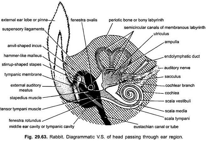
Generally in all mammals a large external ear or pinna or auricle is found which is movable in most animals. It is a skin covered elastic cartilaginous projection from the lateral sides of head. The pinna is a wide-mouthed funnel usually capable of being moved or turned in different directions by its muscles.
The opening of the funnel-shaped pinna leads into a tubular passage, called the external auditory meatus. It ends into a cone-shaped membrane, called the tympanum or ear drum. The walls of auditory meatus are covered by skin containing hairs, oil glands and wax glands. The hairs, oil and wax protect the ear drum from the harm caused due to dust and small insects.
The odour of the wax inhibits the insects to enter the canal. The function of the external ear is to collect the sound vibrations and to detect the direction of sound vibrations and send them to middle ear.
ii. Middle Ear:
The middle ear is the tympanic cavity which starts from the tympanic membrane or ear drum. It is an air-filled cavity enclosed in the tympanic bone. The ear drum is exposed to the impact of sound vibrations caused due to the sound in air. The tympanic cavity is connected with the pharynx by a tubular passage, the Eustachian canal (named after Bartolommeo Eustachio) for equalising the air pressure on both the sides of the ear drum.
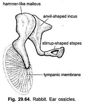
The tympanic cavity is closed internally by a wall which has two windows, opening into the internal ear. These are the upper oval fenestra ovalis and the lower rounded fenestra rotunda. The mammalian middle ear is characterised by the presence of a chain of three tiny bones, the ear- ossicles, extending from the tympanic membrane to the fenestra ovalis.
These ear-ossicles from outside are hammer-shaped malleus, anvil- shaped incus and stirrup-shaped stapes. These bones represent the articular, quadrate and hyomandibular bones of other vertebrates. The ear-ossicles take up the sound vibrations from the tympanic membrane and convey them to the internal ear.
iii. Internal Ear:
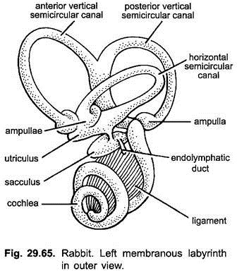
The internal ear is a well- developed and complicated structure, called the membranous labyrinth. It is enclosed in a bony auditory capsule of the same shape as the membranous labyrinth, formed by the periotic bone. The space between the bony capsule and the membranous labyrinth is filled with a liquid, called perilymph.
A similar fluid is found within the membranous labyrinth, called the endolymph having tiny calcareous particles, the otoliths. The membranous labyrinth is formed of a larger dorsal utriculus and a smaller ventral sacculus forming the body proper, and semicircular canals and cochlea.
(a) Utriculus and Sacculus:
The utriculus and sacculus are connected together by a small duct, called sacculo-utricular canal. A small, narrow endolymphatic duct arises from the sacculus which ends blindly against the cranium into an endolymphatic sac. From the sacculus arises a spirally coiled tube called cochlear duct or lagena, which is well developed in rabbit and other mammals.
Both the utriculus and sacculus have a special group of sensory cells called the macula. These cells bear five projecting hairs which are embedded in jelly containing otoliths. Macula of utriculus and salcculus are called macula utriculi and macula sacculi respectively. The bony labyrinth around the utriculus and sacculus is called the vestibule.
(b) Semicircular Canals:
Three semicircular canals connect at both the ends with utriculus. These are external, anterior and posterior semicircular canals, situated at right angles to each other. The anterior and posterior semicircular canals arise from a common canal, after their origin from utriculus are called crus commune. Each semicircular canal has a swollen ampulla at its lower end.
Each ampulla has a sensory area known crista ampullaris which is formed by sensory hair cells and supporting cells. The hairs are embedded in a jelly cone having no otoliths.
The VIII cranial or auditory nerve supporting to ear divides into two branches, vestibular branch supplying to the semicircular canals, utriculus and sacculus and a cochlear branch supplying to the cochlea.
(c) Cochlea:
The spirally coiled watch spring-like cochlear duct or lagena arising from sacculus is enclosed within the similarly spirally coiled cochlear canal of the periotic bone, which together constitute the cochlea. It is the organ of hearing.
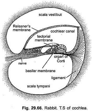
Thus, the cochlea is a bony tube lined with connective tissue. In cross section the cochlea shows three chambers or canals- Middle chamber or scala media is the cochlear duct arising from the sacculus and is filled with endolymph. It terminates blindly at the apex of spiral or cochlea. The upper and lower canals of the cochlea are scala vestibuli and scala tympani respectively.
These are parts of bony labyrinth or cochlear canal and are filled with perilymph. Both the canals are longitudinally separated from each other by a spiral lamina but communicate with each other at the tip of spiral by a small opening, called the helicotrema. The scala vestibuli and scala tympani communicate basally with the fenestra ovalis and fenestra rotunda respectively.
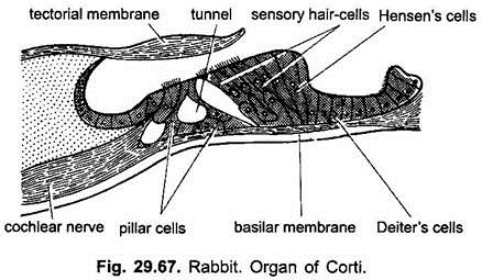
The epithelial wall of the scala media rests above on the Reissner’s membrane and below on the basilar membrane. Basillar membrane is formed of tightly stretched transverse connective tissue fibres. It contains a series of sensory receptor hair cells and long columnar supporting cells.
Hair cells are basally connected with the nerve fibres of cochlear nerve of auditory nerve. Over the hair cells touching the hairs is found a thin, gelatinous, ribbon-shaped sheet of connective tissue, called the tectorial membrane. Both of these are collectively called the organ of Corti.
Working of Ear:
Ear performs two functions- hearing and equilibrium. The cochlear duct of sacculus of the membranous labyrinth is responsible for hearing, while maculi of sacculus, utriculus and cristae of semicircular canals help in equilibrium.
Hearing:
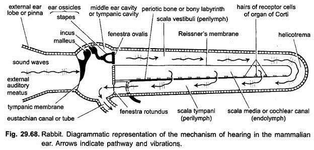
The sound waves are collected by the movable pinna which travel through the external auditory meatus and cause the ear drum to vibrate. The vibrations are then transmitted through the ear-ossicles of the middle ear and fenestra ovalis into the perilymph of internal ear.
The vibrations of the membrane of fenestra ovalis cause alternate increase and decrease in the pressure of the perilymph of scala vestibuli which is transmitted to the scala tympani through helicotrema and escape through the fenestra rotunda back into the middle ear. The membrane of fenestra rotunda due to the pressure bulges out into the middle ear. Thus, it acts as a pressure-relief valve.
The vibrations of the perilymph of scala tympani and scala vestibuli cause endolymph of the scala media and basilar membrane to vibrate. These vibrations cause the tectorial membrane floating in the endolymph of scala media to brush the sensory hairs of organ of Corti. Finally the stimulated hair cells of the organ of Corti send a message through nerve impulses which are carried by the VIII cranial nerve to the brain. Such impulses are interpreted as sound by the brain.
Equilibrium:
The sensory patches of the ampulla of semicircular canals called cristae and utriculus and sacculus are called maculae are responsible for maintaining equilibrium of the body.
Any change in the equilibrium (orientation) of the body stimulates the hair cells of the cristae and maculae due to movement in endolymph and otoliths in them. Maculae respond to the change in the posture of head and body. While the cristae respond to the changes in the direction or rotational movements of head. Cristae lack otoliths.
