The following points highlight the three main types of skeleton found in animals. The types are: 1. Exoskeleton 2. Hydro-Skeleton 3. Endoskeleton.
1. Exoskeleton:
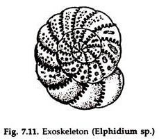
Hard structures derived from epidermis or dermis, present outside the body and partly or almost fully enclosing it are called exoskeleton. Many fresh water protozoon secrete a chitinoid (Arcella), calcic (Elphidium) shell (Fig. 7.11); some protozoon living in damp soil secrete a cementing material in which sand and soil particles are embedded and acts as a shell.
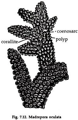
The polyps of stony corals secrete calcium carbonate, which forms a skeleton beneath and around them. With the growth and multiplication of polyps, new materials are added and the coral grows in size (Fig. 7.12). The corals may be flat, upright and branched or of any other shape. The chief constituent of the coral reef is the exoskeleton of stony corals of many generations.
In annelids, the exoskeleton is a thin cuticle or chitin around the body and bears a number of pores for communication of internal organs with the exterior. The exoskeleton in arthropods is a complex glycoprotein secreted by the epidermis. It encloses the body almost completely (Fig. 7.12).
It is not a complete tube but thick pieces are joined together to allow free movement. In appendages, the exoskeleton is tubular. The skeleton may extend inwards to line parts of the gut (crustacea) or trachaea (insects).
The hard covering made of several pieces (Chiton), two valves (mussels) or a single piece (gastropods) enclosing the body is secreted by the mantle in molluscs. The exoskeleton, however, is absent in slugs (gastropod: pulmonate) and cephalopods. The chief constituent of molluscan shell is calcium carbonate lined internally with a layer of nacre or mother of pearl and externally by a layer of horny protein.
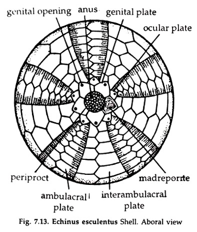
In echinoderms, the body in most cases, is protected by a hard exoskeleton (Fig. 7.13), except holothuroids. The skeleton is composed of ossicles of calcium carbonate of various shapes and sizes bound together by connective tissue. Chitinoid spines, wart and tubercles are present in the ossicles. In sea urchin, the exoskeleton forms a test, bearing many perforations and spines of varying length and thickness.
ADVERTISEMENTS:
In holothuroids, the exoskeleton is represented by a thin cuticle covering the body and numerous small ossicles in the dermis and the two together form a tough skin.
2. Hydro-Skeleton:
The hydro skeleton is not that important in higher forms, but it has useful functions in lowly organised animals and in larval forms. The fluid in the body under pressure—helping in locomotion, is considered as hydro skeleton. The coelomic fluid in earthworm, the haemocoelomic fluid in the foot of arthropods, molluscs and podocyst of mollusc larvae, the notochord in Branchiostoma are considered as hydro skeleton.
ADVERTISEMENTS:
Contraction of circular and longitudinal muscles in the body wall of an earthworm exerts pressure on the coelomic fluid and forces the animal to elongate or shorten (widen). Increased pressure on the haemocoelomic fluid (blood) in the foot due to contraction of foot muscle in mussels, forces the foot to elongate and it is pushed forward into the substratum.
A sudden rush of haemocoelomic fluid causes forceful extension of the legs during a jump in jumping spiders. The notochord in Branchiostoma is an elongated cord of cells filled with a fluid. The firm connective tissue sheath surrounding it helps to maintain the turgidity. The notochord is flexible but firm and prevent shortening of the body during movement, due to contraction of the longitudinal segmental muscles.
3. Endoskeleton:
The skeletal structures present in the body wall or in deep body tissue are collectively known as endoskeleton.
Invertebrate Endoskeleton:
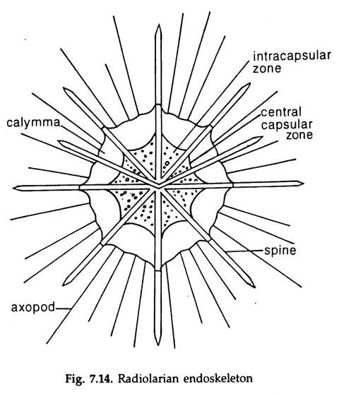
Radiolarians, a group of large planktonic protozoa, have a very delicate endoskeleton made of elaborate silica lattices (Fig. 7.14). Variously shaped spicules made of sponging fibres or calcium carbonate or both present in the body, support the sponges and help to maintain shape. Portions of some of the spicules project beyond body surface.
In cephalopods, internal shell of calcareous particles arranged in the form of lamellae and a cartilaginous cranium enclosing the nerve ganglia are present. The ossicles made of calcium carbonate, situated in the dermis of echinoderms are considered as endoskeleton by some zoologists.
Vertebrate Endoskeleton:
The endoskeleton of vertebrates is made up of bones and cartilages, the latter contributing only a small fraction. The entire endoskeleton of Agnatha (jawless) and Chondrichthyes (sharks) is made of cartilage. The chief constituents of bones is calcium phosphate.
ADVERTISEMENTS:
Vertebrate bones develop from mesoderm cells, and are of two types— the membrane bone (intramembranous ossification) and cartilage bone or long bone (intra-cartilaginous ossification).
In both the cases, prior to deposition of bony or cartilaginous materials, the mesoderm cells are transformed into mesenchyme’s. In the membrane bone, the materials contributing to the formation of bone are deposited directly in between the cells and around blood vessels.
In the long bone, in the foetal stage, a part of condensed mesenchyme is replaced by hyaline cartilage. In the adult, the cartilage is replaced by bone. The membrane bones are usually flat and light but quite strong, and present in the roof and sides of the skull. The long bones are thick and heavy.
The vertebrate skeleton is divided into a somatic skeleton (Gr. soma = body), present in the body wall and visceral skeleton (Gr. viscera = bowels) restricted to primitive pharyngeal wall and gills. The somatic skeleton is subdivided into an axial skeleton, comprising vertebral column, sternum, ribs and most of the skull and an appendicular skeleton consists of girdles, both pectoral and pelvic and limb bones.


