In this article we will discuss about:- 1. Shell of the Apple Snail 2. External Features of Apple Snail 3. Torsion 4. Locomotion 5. Pallial Complex 6. Body Cavity 7. Digestive System 8. Respiratory System 9. Circulatory System 10. Excretory System 11. Nervous System 12. Reproductive System 13. Fertilization and Egg Laying in Apple Snail 14. Development Fertilization and Development in Apple Snail.
Contents:
- Shell of the Apple Snail
- External Features of the Apple Snail
- Torsion of the Apple Snail
- Locomotion in the Apple Snail
- Pallial Complex in the Apple Snail
- Body Cavity of the Apple Snail
- Digestive System of the Apple Snail
- Respiratory System of the Apple Snail
- Circulatory System of the Apple Snail
- Excretory System of the Apple Snail
- Nervous System of the Apple Snail
- Reproductive System of the Apple Snail
- Fertilization and Egg Laying in Apple Snail
- Development Fertilization and Development in Apple Snail
1
. Shell of the Apple Snail:
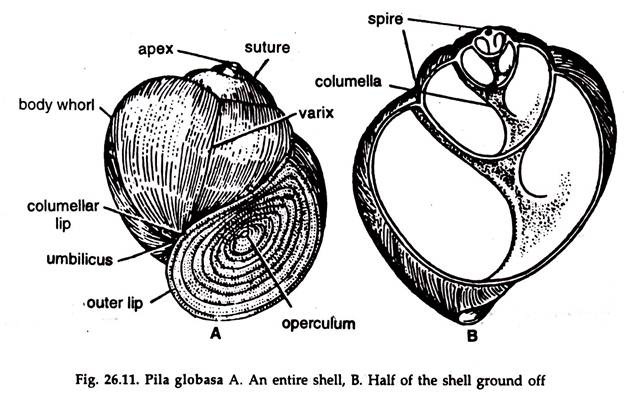
1. The body is enclosed in a thick, spirally coiled, dextral globular shell.
ADVERTISEMENTS:
2. The shell is a broad cone, coiled round a central axis, the columella, in a spiral manner.
3. The top of the shell is called apex.
4. Starting from the apex, the whorls are larger in size, the last or the body whorl being the largest (Fig. 26.11).
5. The whorl next to the body whorl is penultimate whorl.
ADVERTISEMENTS:
6. The junction of two successive whorls is known as suture.
7. A large aperture called mouth is present in the body whorl. The margin of the aperture is smooth and known as peristome.
8. The aperture can be closed by an operculum which is a flat calcareous plate, lunate-oblong in outline.
ADVERTISEMENTS:
9. Concentric growth rings around the centre or nucleus are present in the operculum.
2. External Features of the Apple Snail:
1. The body is soft, slimy and lodged within the shell.
2. It is attached to the columella, the spirally twisted axis of the shell, by a columellar muscle arising from the foot.
3. The muscle prevents the animal from extending out of the shell beyond a certain limit and also helps to withdraw it into the shell.
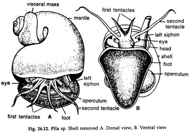
4. The body is divisible into head, foot and visceral mass (Fig. 26.12).
5. The head is well-marked and drawn anteriorly into a snout.
6. The head carries two pairs of tentacles:
i. The shorter pair are anterolateral in position and called labial palps or first tentacles.
ADVERTISEMENTS:
ii. The longer pair or second tentacles are thread-like and bear stalked eyes at the base.
7. Two fleshy projections called nuchal lobes or pseudopipodia are seen on the two sides of the head. The left nuchal lobe is highly developed and form the respiratory siphon.
8. The mouth is a vertical slit at the end of the snout. True lips are absent. The plicate edges serve as lips.
9. The anus is a small round aperture. It is anteriorly placed at about 6 mm distance from the right pseudopipodium.
10. The foot is highly muscular and more or less triangular in shape. If viewed ventrally, the sole or the creeping surface is flat.
11. The operculum is attached to the dorsal surface of the posterior part of the foot.
11. The skin covering the visceral mass is known as the pallium or mantle.
12. In a retracted state, the mantle forms a cloak over the anterior part of the body including the head and its appendages.
3. Torsion of the Apple Snail:
The twisting of the visceral mass on head- foot axis is termed torsion, a phenomenon peculiar to gastropod molluscs. The gastropod in the early embryonic stage is bilaterally symmetrical.
In the process of torsion, the head-foot axis remains unchanged, the visceral hump of the embryo rotates 180°, shifting the mantle cavity and the posterior part of the visceral mass forward and upwardly, bringing the anus close to the mouth.
The torsion is usually due to unequal growth of the two halves of the visceral mass associated with muscular contraction or rarely by muscular contraction alone. The active growth of one side shifts the structures towards the opposite side, the growth of the structures in the latter half being suspended, viz. the active growth of the left side shifts the structures towards the right and vice versa.
The clockwise twisting is called dextral and the anticlockwise sinistral. In dextral forms, the shell mouth (opening of the shell) is to the left and the twisting of the shell is clockwise, when the shell is held with the apex away from the observer and the mouth ventral. In sinistral forms the situation is reverse. The majority of the gastropods are dextral e.g. Pila, Achatina, etc.
4. Locomotion in the Apple Snail:
Locomotion in Pila is slow and effected by crawling on the surface with the help of a more or less triangular foot with a flat sole.
1. The foot is provided with vertical, longitudinal and transverse muscles.
2. Glands in the foot secrete a copious amount of slimy mucus, making the surface on which-the snail moves, slippery.
3. The foot is protruded through the mouth of the shell due to influx of haemolymph in the pedal sinus.
4. Contraction of the transverse muscles forces the haemolymph forward resulting in the forward elongation of the foot.
5. Contraction of vertical muscles creates wave-like movement on the sole of the foot, helping it to move forward.
6. Contraction of longitudinal muscles pull the posterior end of the foot forward and the snail creeps slowly on the substratum.
5. Pallial Complex in the Apple Snail:
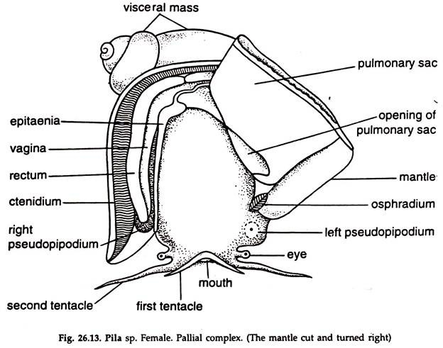
The structures present in the mantle or pallial cavity are collectively called pallial complex. The body is enclosed in a mantle or pallium. In the body whorl it forms a cloak over the body, enclosing the mantle or pallial cavity (Fig. 26.13).
In the rest, the pallium is thin and adherent to the visceral mass. The opening of the mantle cavity is anterior and wide. A longitudinal ridge, the epithelium, incompletely divides the cavity into a right and left chamber. The right or branchial chamber contains ctenidium, rectum and genital duct. The left or pulmonary chamber contains the pulmonary sac.
6. Body Cavity of the Apple Snail:
In the adult, the coelom is greatly reduced and restricted to pericardium (pericardial cavity) and lumen of the kidney (urocoel). The haemocael acts as channels for haemolymph circulation.
7. Digestive System of the Apple Snail:
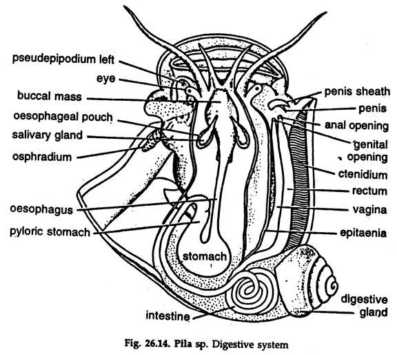
The digestive system consists of a coiled alimentary canal, a pair of salivary glands, a pair of oesophageal pouches and a bilobed digestive gland (Fig. 26.14).
Alimentary Canal:
It is divided into three zones—the foregut, midgut and hindgut:
The fore and hindgut are of stomodaeal and proctodaeal origin, respectively and lined by ectoderm cells, while the midgut is arch enteric and lined by endoderm cells.
i. Foregut:
It includes mouth, buccal mass and oesophagus:
1. Mouth:
The mouth is a slightly ventral, vertical slit at the tip of the snout. The first pair of tentacles, short in length, are anterolateral projections of the snout and considered as labial palps. The mouth leads into a thick- walled buccal cavity.
2. Buccal mass:
It is a fairly large pyriform muscular structure. The buccal cavity is a spacious chamber, major portion of which is occupied by an odontophore. Anteriorly, the buccal mass bears two dorsolaterally placed horny jaws, the anterior cutting edge of which bears two or three teeth. The buccal cavity opens into the oesophagus.
Odontophore:
A thick-walled muscular structure.
It consists of a radula, radular muscles, a pair of sub-radular cartilages and a sub-radular organ with a radular sac:
a. Radula:
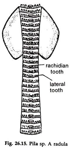
It is a thin, spoon-shaped, chitinoid ribbon (Fig. 26.15), the narrow median strip of the broad anterior part is exposed to the floor of the buccal cavity and the narrow posterior portion is lodged in the radular sac.
i. The radula is movably placed on a pair of radular cartilages operated by large radular muscles, the two constituting major part of the odontophore.
ii. Numerous transverse rows of teeth are present on the radular ribbon. The teeth in a transverse row are one median or rachidian, one lateral and two marginal, the radular formula being 2. 1. 1. 1. 2.
iii. Distally, the radular teeth bear broad cutting edge or cusp. A rachidian tooth has a median and two or three lateral cusps on each side.
3. Oesophagus:
A long median tube, narrows posteriorly, runs medially backwards, turns left and enters the visceral mass to join the stomach. The salivary glands open into the dorsal aspect of the buccal cavity close to the origin of the oesophagus and the oesophageal pouches (glands) open close to the openings of the salivary glands.
ii. Midgut:
The stomach constitutes the midgut.
Stomach:
A somewhat oval-shaped, dark red, muscular sac embedded partially in the digestive gland. It has two chambers. The stomach with the oesophagus and the intestine is a U-shaped body. The base of the U is formed by the stomach and the arms by the oesophagus and the intestine.
iii. Hindgut:
It is a long, narrow tube and divisible into intestine and rectum:
Caecum:
A small diverticulum at the origin of the intestine, not clearly discernible from outside.
Intestine:
A narrow tube, forms 2½ to 3 coils in the mass of the digestive gland. Emerging from the gland it enters the mantle cavity and joins the rectum.
Rectum:
A wide tube, exposed to the mantle cavity and adherent to the inner surface of the mantle on the right side, parallel to the ctenidium. It runs anteriorly to open through the anus in the right side of the mantle cavity near its opening.
Digestive Glands:
The salivary glands, oesophageal pouches (glands) and digestive gland constitute the digestive glands:
1. Salivary glands:
A pair of whitish glands, one on either side of the oesophagus. The glands are alveolar type and open into the dorsal aspect of the buccal cavity through narrow ducts.
2. Oesophageal pouches (glands):
A pair of sac-like glands, placed laterally at the origin of the oesophagus and open into the buccal cavity close to the opening of the salivary glands.
3. Digestive gland:
A large, bilobed, brownish or dull green mass. Roughly, it is a spirally coiled, triangular plate. Two ducts from two lobes of the gland unite to form a common duct before opening in the stomach.
Feeding and Digestion:
Pila feeds upon plant matters.
i. The labial palps help in bringing the food close to the mouth.
ii. Small pieces of plant materials may be cut by the horny jaws and passed to the buccal cavity through the mouth.
iii. The major part of the food is procured by rasping with radula. The food is held tightly by the labial palps and the anterior margin of the musculature forming the floor of the buccal mass.
iv. The radula is protruded out through the mouth by the action of protractor muscles. The alternate action of upper and lower sets of muscles cause forward and backward, a chain-saw like movement of the radula and the food matter is rasped by the cusps of the radular teeth and passed to the oesophagus.
v. The cellulosic cell walls of a considerable portion of the plant food are broken mechanically during rasping and the contents of the cells released, which facilitates digestion.
vi. The food gets mixed with saliva and secretion of the oesophageal pouches in the buccal cavity, before it is passed to the oesophagus. In the stomach the food is mixed with the secretion of the digestive gland. Extracellular digestion is partial in the stomach.
vii. Small particles of food pass to the digestive gland through the digestive duct and intracellular digestion completes the hydrolysis of food matters in the digestive gland.
viii. The undigested part of the food pass out in the form of strings through the anus.
8. Respiratory System of the Apple Snail:
Pila are remarkable amphibious inhabitants of fresh waters in tropical countries. It is noted for its ability to respire both under water and in air, being provided with a gill and a pulmonary sac. When under water,
Pila maintains the usual respiratory current through the right side of the mantle cavity which contains the gill; at intervals it crawls to the surface or swims up by lashing the snout and tentacles, and forms a funnel leading into the pulmonary sac by rolling up the left nuchal lobe. Pila globosa is able to make long excursions on land.
The respiratory organs consist of a gill, a pulmonary sac and the right and left nuchal lobes and afford both terrestrial and aquatic respiration. The pallial cavity is incompletely partitioned by a fleshy fold.
The epitaenia into two chambers, the right one branchial chamber containing the gill and the left one pulmonary chamber containing the pulmonary sac:
a. Gill Respiration:
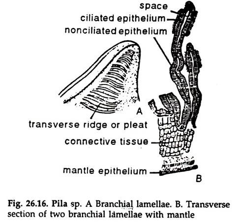
1. The gill or ctenidium consists of a series of thin triangular leaflets or lamellae. The lamellae are largest in the middle but become smaller towards the extremities (Fig. 26.16) and are lodged in the right portion of the mantle cavity. Each lamella bears a varying number of transverse ridges or pleats on its both outer and posterior surfaces.
2. The lamellae are attached to the mantle along an axis known as ctenidial axis. The attachment is effected by the broad bases of the lamellae, the narrow apices hanging freely in the mantle cavity.
Mechanism of Respiration:
Due to extension of the mantle cavity and nuchal lobes water enters the mantle cavity through the left lobe or siphon, which forms a distinct gutter.. In its course, water comes in contact with the osphradium. Bathing the gill, the water goes out through the right nuchal lobe or siphon due to the contraction of the mantle cavity. The gill filaments are highly vascular and gaseous exchange takes place there.
b. Pulmonary respiration:
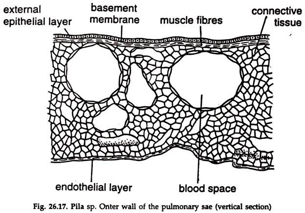
1. The pulmonary sac hangs down like a sac from the roof of the pulmonary chamber (Fig. 26.17). The sac opens to the exterior at the extreme posterior limit of the mantle cavity.
Mechanism of Respiration:
When out of water, air enters the pulmonary chamber of the mantle cavity through its opening. With the contraction of the muscles in the wall, the cavity of the pulmonary sac enlarges and air is snacked into it.
Gaseous exchange takes place in the highly vascularized wall of the pulmonary sac. Siphon formed by the left nuchal lobe does not play any role in it. The inspired air is forced out by the compression of the pulmonary sac due to relaxation muscles in its wall.
9. Circulatory System of the Apple Snail:
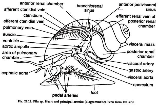
The circulatory system in Pila is open type and consists of heart, pericardium, arteries, sinuses and veins (Fig. 26.18):
Phagocytic amoebocytes are present in the haemolymph. The haemolymph contains dissolved haemocyanin, the respiratory pigment, a compound of copper and protein.
i. Pericardium:
1. A thin-walled, ovoid sac lying dorsally on the left side of the body whorl, behind the mantle cavity and extending anteriorly up to the stomach and digestive gland.
2. The pericardium encloses the heart, main aortic arches and aortic ampulla.
ii. Heart:
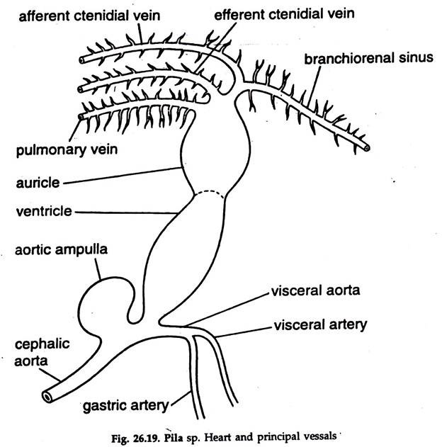
The heart has a single auricle and a ventricle (Fig. 26.19):
1. Auricle:
A thin-walled, highly contractile, triangular sac.
It receives:
a. An efferent ctenidial vein from ctenidium.
b. An efferent renal vein at its apex, from the posterior chamber of the renal organ.
c. A pulmonary vein at a slightly lower level at the anterior end, from the pulmonary sac.
The auricle communicates with the ventricle by an auriculoventricular opening, guarded by two semilunar valves, opening towards the ventricle.
2. Ventricle:
A thick, spongy, ovoid chamber. It sends an aortic trunk from its lower end, which divides immediately into two branches—cephalic and visceral aorta:
The opening between the aortic trunk and ventricle is guarded by two semilunar valves, which prevent backflow of haemolymph to the ventricle.
iii. Arteries:
a. Cephalic aorta:
It bears a bulbous outgrowth, the aortic ampulla, which controls haemolymph pressure. The aorta proceeds anteriorly and sends branches from both the outer and inner sides.
Branches from the outer side:
i. Cutaneous artery to skin.
ii. Oesophageal artery to oesophagus.
iii. Pallial artery to organs of the left half of the mantle cavity and osphradium.
Branches from the inner side:
iv. Pericardial artery to pericardium.
v. Renal artery to posterior renal chamber.
The cephalic aorta proceeds anteriorly along the left side of the oesophagus for some distance, then crosses to the right and sends many branches, the important ones are:
vi. Right pallial artery to right side of the mantle.
vii. Right siphonal artery to right siphon.
viii. Penial artery to penis.
ix. Optic arteries to eyes.
x. Tentacular arteries to tentacles
xi. Radular artery to radular sac.
xii. Pedal artery to foot.
b. Visceral aorta:
It runs backwards into the visceral mass and supplies haemolymph to visceral organs.
The important branches are:
i. Visceral artery to digestive gland.
ii. Gastric artery to stomach.
iii. Intestinal artery to intestine.
iv. Hepatic artery to digestive gland and gonad.
The aorta terminates in the rectum.
iv. Sinuses:
The haemolymph from the finer branches of the arteries passes into small lacunae. Many small lacunae unite to form a large sinus.
There are four large sinuses:
a. Anterior perivisceral sinus:
It lies above the foot and beneath the floor of the pallial cavity.
b. Anterior periintestinal sinus:
Located in the columellar axis, runs along the coil of the intestine up to the junction of the anterior and posterior renal chambers.
c. Banchiorenal sinus:
It lies along the right side of the anterior renal chamber.
d. Pulmonary sinus:
It is adherent to the walls of the pulmonary sac.
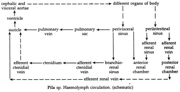
The haemolymph finally passes to the perivisceral and periintestinal sinuses and carried to auricle by veins directly or through gills, mantle and kidney.
v. Veins:
a. Afferent ctenidial vein:
It lies above the rectum and receives branches from rectum, terminal part of the genital duct, perivisceral sinus and branchiorenal sinus and sends haemolymph to the gill lamellae.
b. Efferent ctenidial vein:
Runs along the roof of the anterior renal chamber and drains haemolymph from gill lamellae, mantle and sends it to auricle.
c. Afferent renal vein:
Located on the roof of the’ posterior renal chamber. It originates from the per intestinal sinus and sends haemolymph to the posterior renal chamber.
d. Efferent renal vein:
Carries haemolymph from posterior renal chamber to the auricle.
e. Pulmonary vein:
Collects haemolymph from the pulmonary sac and open into the auricle. The efferent ctenidial vein and pulmonary vein carry oxygenated haemolymph, the afferent renal vein carries deoxygenated haemolymph to the auricle.
10. Excretory System of the Apple Snail:
The kidney is the chief excretory or renal organ:
Kidney:
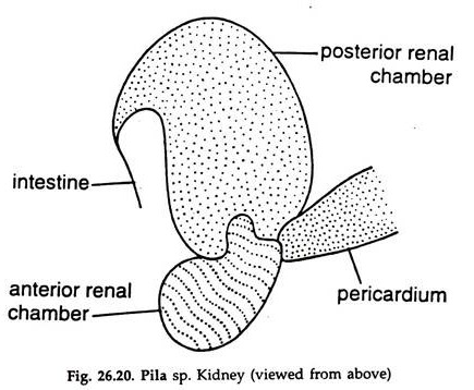
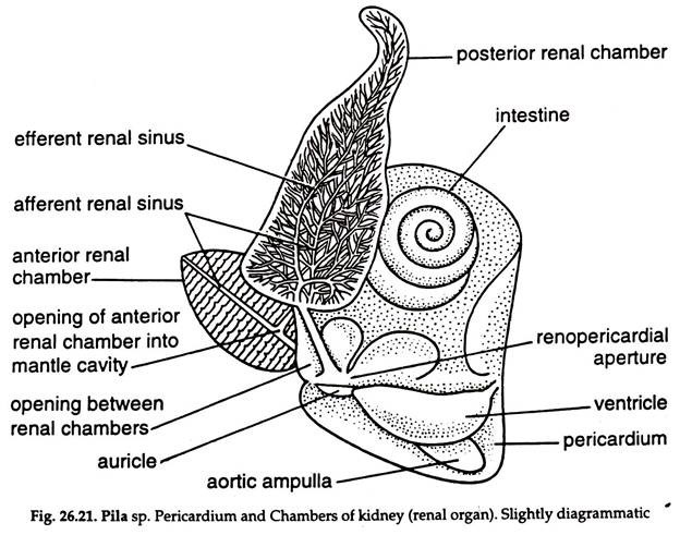
The kidney is a large, thin-walled sac lying behind the pericardium. It consists of two distinct renal chambers, a right anterior and a left posterior (Figs. 26.20, 26.21):
1. Anterior chamber:
a. It is an ovoid, reddish organ situated in front of the pericardium and posterior renal chamber.
b. The anterior renal chamber projects into the mantle cavity and opens into it in a deep crypt through a slit-like opening, the renal pore, lying to the right of the epithelium.
c. The lamellae on the roof of the chamber are arranged on either side of a median longitudinal axis, the efferent renal sinus.
d. The lamellae on the floor are similarly arranged on either side of a median axis, the afferent renal sinus.
2. Posterior chamber:
a. It is a broad, brownish, hook-shaped chamber situated behind the anterior renal chamber in between rectum on the left and pericardium and digestive gland on the right. It possesses a large cavity.
b. The roof of the chamber is richly supplied with haemolymph.
c. The posterior chamber communicates with the pericardium by a Reno pericardial aperture at one end and with the anterior renal chamber at the other end.
Digestive gland also plays a role in excretion. The lobules contain excretory cells which eliminate waste materials.
The nature of nitrogenous wastes in Pila varies with season and habitat. Ammonia, urea and uric acid are major constituents of nitrogenous wastes. Pila living in water during aquatic phase produces ammonia, while living out of water during terrestrial phase it excretes urea and uric acid, depending on the period of land life.
11. Nervous System of the Apple Snail:
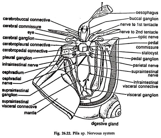
A central nervous system, a peripheral nervous system and a visceral nervous system constitute the nervous system. (Fig. 26.22)
The central nervous system comprises paired cerebral, buccal, pleural, pedal, visceral ganglia and an impaired supraintestinal ganglion; cerebral, buccal, pedal commissures; paired cerebrobuccal, cerebropleural, cerebropedal connectives; supra and infraintestinal visceral connectives and supra and infraintestinal nerves.
Nerves arising from the ganglia and connectives constitute the peripheral nervous system. Nerves innervating some of the visceral organs form visceral nervous system.
Cerebral ganglia:
The cerebral ganglia are paired roughly triangular in shape, situated anteriorly on the dorsolateral sides of the buccal mass at its anterior end. Nerves arising from the cerebral ganglia innervate the snout, tentacles, eyes and statocysts.
Cerebral commissure:
The two cerebral ganglia are connected by a thick, band-like commissure medially, located dorsal to the buccal mass.
Labial commissure:
A fine commissure, connects the two cerebral ganglia ventrally.
Buccal Ganglia:
Two small, oval, roughly triangular, ganglia embedded in the dorsolateral sides of the buccal mass at its junction with the oesophagus, near the radular sac.
Nerves arising from the buccal ganglia innervate the buccal mass, salivary glands and oesophagus.
Buccal commissure:
A slender nerve connecting the two buccal ganglia, located ventral to the oesophagus. Cerebrobuccal connectives
Each buccal ganglion is connected with the cerebral ganglion of the side by a slender cerebrobuccal connective.
Pleuropedal Ganglionic Mass:
A pair of ganglionic masses, one on either lateral side, posteroventral to the buccal mass. The pleural and pedal gangila of each side are more or less fused, only a slight notch separating the two. The sub-intestinal ganglion fuses with the right pleurepedal ganglionic mass. Pleural ganglia send nerves to the mantle complex and nuchal lobes and adjacent organs and the pedals to the foot.
Cerebropleural connectives:
Two Each runs posteroventrally from the cerebral ganglion, around the oesophagus and joins the pleural region of the pleuropedal ganglion of the side. Its origin is medial to that of the cerebropedal connective.
Cerebropedal connectives:
A pair each runs posteroventrally from the cerebral ganglion, around the oesophagus and joins the pedal region of the pleuropedal ganglion of the side.
Pedal Commissure:
A thick nerve connecting the pedal regions of the two pleuropedal ganglia.
Infraintestinal nerve:
A slender nerve, close to the pedal commissure, joining the pleural regions of the two pleuropedal ganglia.
Visceral ganglion;
It is formed by the fusion of two spindle-shaped ganglia and situated close to the posterior border of the mantle cavity at the right of the pericardium. The ganglion sends nerves to kidney, heart, digestive gland, intestine and reproductive organs.
Supraintestinal visceral connective:
Running backwards from the pleural part of the left pleuropedal ganglia, joins the visceral ganglion. It bears a supraintestinal ganglion at about its middle.
Infraintestinal visceral connective:
Runs backward from the pleural part of the right pleuropedal ganglia and joins the visceral ganglion. The supra and infraintestinal visceral connectives are also designated as left and right pleurovisceral connective, respectively. Both of them send nerves to adjacent organs.
Supraintestinal nerve:
A slender nerve runs obliquely backwards and connects the right pleuropedal ganglia with the supraintestinal ganglion.
Supraintestinal ganglion:
It is almost fusiform and situated slightly posterior to the pleuropedal mass at some distance from it on the supraintestinal visceral or left pleurovisceral connective. The ganglion sends nerves to gill, pulmonary sac, etc.
Receptors and Sense Organs in the Apple Snail:
The sense organs consist of tactile organs, osphradium, statocysts and eyes:
Tactile organs:
Tactile sensory cells are scattered all over the exposed body surface. In addition, the labial palps and second pair of tentacles are tactile organs.
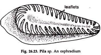
Osphradium:
Single, a small body suspended ventrally from the anterior border of the mantle cavity, on the left side (Fig. 26.23). Osphradium is test organ for water and food.
Statocysts:
Two cream-white sacs, each located posterior to the pedal ganglion, of the pleuropedal ganglionic mass, close to it and kept in position by muscles.
Structure:
a. A pyriform, cream-white, leathery sac.
b. The internal lining epithelium contains sense cells.
c. A statolith is present in the cavity. The statocysts are balance organs and innervated by cerebral ganglia.
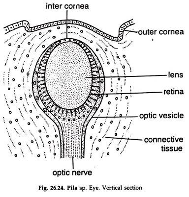
Eyes:
Two, borne on short stalks at the base of the second pair of tentacles:
a. The outermost layer of an eye is an optic vesicle, formed by a thick layer of fibrous connective tissue (Fig. 26. 24).
b. The optic vesicle is open at the tip and covered by transparent outer and inner cornea.
i. The outer cornea is a continuation of the epithelium of the eye stalk, the cells are small, squarish and transparent.
ii. The inner cornea is also transparent and continuation of the connective tissue around the optic vesicle.
c. The inner lining of the optic vesicle is retina, made of two types of cells:
i. Visual cells:
Large, broad and with brush border.
ii. Packing cells:
Small, wedged between visual cells.
d. The space enclosed by the retina is occupied by an ovoid lens.
e. The optic nerve enters the optic vesicle at the posterior end.
The eyes of Pila are poor photoreceptors.
12. Reproductive System of the Apple Snail:
The sexes are separate, sexual dimorphism is not pronounced. The gonad, testis or ovary is single and occupies the same position in male and female. It is restricted to about half the height of the inner concave surface of the first two to three whorls of the digestive gland. The gonad is covered by the mantle, which is very thin and transparent.
Male reproductive system:
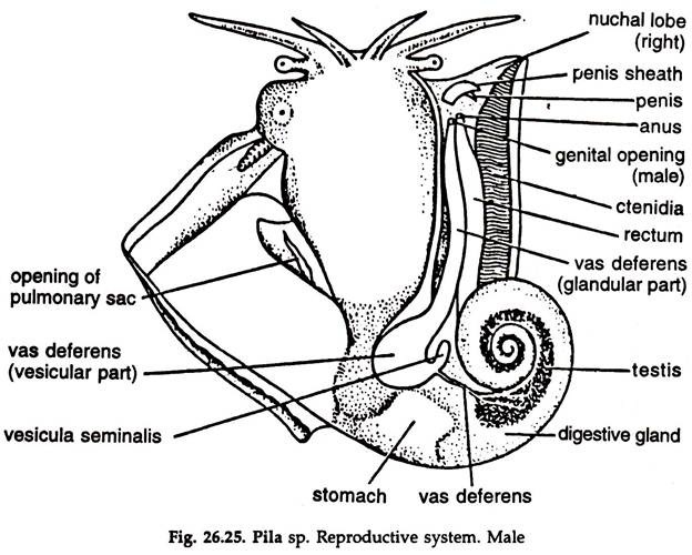
It consists of a testis, a vas deferens and a penis. Testis Much branched, cream coloured and clearly visible through the transparent mantle. It enlarges posteriorly. The lobes of the testis are exposed after the removal of the mantle (26. 25).
Vas deferens:
Many fine ducts, the vasa efferentia, emerging from the lobes of the testis join to form the vas deferens, which leaves the testis at the posterior end and runs along the inner border of the posterior renal chamber. The vas deferens has three distinct zones.
a. Tubular part:
The slender first part runs along the inner edge of the posterior renal chamber.
b. Glandular part:
Terminal part of the Vas deferens. It is fairly large, swollen at the beginning and narrows distally, being lodged in the mantle cavity, parallel to the rectum and opens at the tip of the genital papilla.
c. Seminal vesicle:
A curved, blind tube at the junction of the tubular and glandular part of the vas deferens.
Penis:
A long, whip-like flagellum, partially en- sheathed; an outgrowth of the mantle, located on the right side of the mantle cavity.
Female Reproductive System:
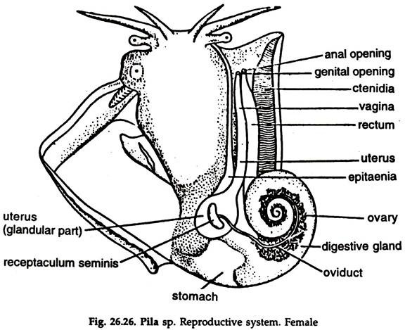
It consists of an ovary and the oviduct with vagina.
Ovary:
Orange-red in colour, smaller than the testis and occupies the same position as the testis in the male (Fig. 26. 26).
Oviduct:
Formed by the joining of a number of narrow ducts from the lobes of the ovary and leaves it at the posterior end.
The oviduct is divisible into two distinct zones:
a. Tubular part:
The slender first part running along the border of the digestive gland, turns downwards above the level of the kidney and joins the bean-shaped semiual receptacle at the junction of the tubular and glandular part. The seminal receptacle receives sperms from the male during mating.
b. Glandular part:
It lies in the mantle cavity, parallel to the rectum and divisible into two zones:
i. Uterus:
A bluntly rounded yellowish sac, lying at the junction of the two renal chambers and narrows down to form the vagina.
ii. Vagina:
Tubular, light cream coloured, narrows down anteriorly and opens through a slit-like aperture on the genital papilla.
13. Fertilization and Egg Laying in Apple Snail:
Pila breeds in monsoon.
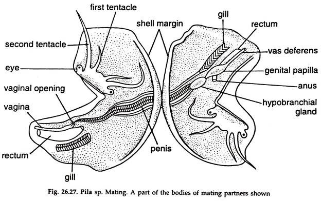
1. Copulation lasts for 2-3 hours. The tip of the penis is inserted into the vagina (Fig. 26.27) and the spermatic fluid is transferred to seminal receptacle, where fertilization occurs.
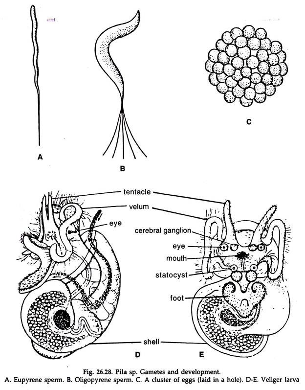
2. The sperm are of two types (Fig. 26.28 A. B), Eupyrene and oligopyrene. The eupyrene type fertilize the ova.
3. The zygote receives albumen and becomes enclosed in a double shell membrane, surrounded externally by a white, calcareous (Fig. 26.28C). egg shell.
4. The embryo remains suspended in the core of fluid albumen in the egg.
5. 200 to 800 eggs, the number being extremely variable in a brood, are laid in sheltered crevices or holes in the soft soil, made by the snout of the mother, above water level. The eggs are held together by a transparent cementing material.
14. Development Fertilization and Development in Apple Snail:
Development starts immediately after fertilization. Development indirect and passes through a veliger stage, which is considered as an advanced larva. Cleavage holobastic and spiral. The gastrula is transformed into veliger, the trochophore stage being suppressed.
In the early stages of development the embryo is bilaterally symmetrical. Owing to asymmetrical growth, the visceral mass twist 180° on the head-foot axis and torsion occurs, leading to the appearance of a veliger larva.
Veliger larva:
The veliger (Fig. 26-28 D. E.) is asymmetrical, transparent, elongate oval in shape and enclosed in a coiled shell for the most part; mantle cavity rudiment is a cleft on the border of the mantle in the right side; foot large; paired tentacles, coelomic sacs, eyes, cerebral ganglia and statocysts are present. The larva metamorphoses to a young pila.