The following points highlight the nine main types of urochordata. The types are: 1. Molgula 2. Botryllus 3. Ascidia 4. Ciona 5. Clavelina 6. Pyrosoma 7. Doliolum 8. Salpa 9. Oikopleura.
Type # 1. Molgula:
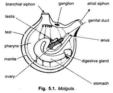
Molgula (Fig. 5.1) is a round, solitary and sessile ascidian, about 2.5 cm long. It is found abundantly in the shallow water of Atlantic and less widely in the Indian Ocean, being fixed to wharfs, piers and rocks. There is no foot. Test is thin and transparent, it is often encrusted with shell pieces and sand. The siphons are well formed, the branchial aperture has 6 lips and the atrial aperture has 4 lips.
In the branchial siphon are 16 branching tentacles. Pharynx is large with narrow stigmata arranged spirally. Dorsal lamina is C-shaped and has no languets. Alimentary canal forms a curved loop.
There is a curved renal vesicle or kidney above the heart. There is a pair of protandrous gonads. Molgula is usually oviparous. The tadpole larva has no ocelli, and in some species there is no tail. Some species of Molgula are viviparous giving birth to a larva which may or may not have a tail in different species.
Type # 2. Botryllus:
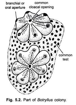
Botryllus (Fig. 5.2) is widely distributed in the Atlantic and the Mediterranean, being found fixed to wharfs, piers and ships in shallow water. It is a colonial ascidian. The test forming a flat encrusting mass with individuals or zooids grouped together in radiating star-shaped fashion. The branchial apertures of zooids are at their outer ends, while their atrial apertures open into a common cloacal chamber opening in the centre of the group of zooids.
The test of all zooids is common forming a gelatinous mass. The structure of each zooids is like that of a simple ascidian. Dorsal lamina has no languets neural gland is dorsal to the brain. Pharynx has rectangular stigmata. There is a pair of hermaphrodite gonads, reproduction is sexual and also by budding, the buds remain permanently in the colony.
Type # 3. Ascidia:
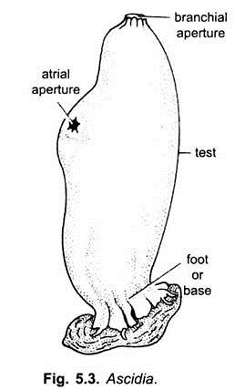
Ascidia (Fig. 5.3) is common in the Atlantic Ocean and Mediterranean sea being fixed to rocks. It is 10 to 12 cm long. The test is thick and cartilaginous. The body is cylindrical with a broad base by which it is fixed. At the free end is a branchial aperture, and some distance from it on one side is an atrial aperture. The siphons are short. The pharynx is large with numerous stigmata but there are no internal longitudinal folds.
ADVERTISEMENTS:
The intestine contains a typhlosole. There is no liver. Dorsal tubercle is horse-shoe-shaped. Dorsal lamina is a longitudinal ridge with no languets. Brain is dorsal to the neural gland. Excretory renal vesicle lies on the stomach and intestine. There is a single hermaphrodite gonad in the intestinal loop.
Type # 4. Ciona:
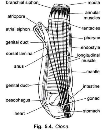
Ciona (Fig. 5.4) is solitary and is found in great abundance on rooks, ships, etc., in cold and temperate seas. It is 10 to 12 cm long with a cylindrical body, It is fixed by a broad foot-like stalk or base. Branchial and atrial siphons are cylindrical, the former being higher up than the latter. The test is thin and transparent. The mantle has longitudinal and circular muscle bands.
The pharynx has no internal folds, the dorsal lamina has languets. Stomach, oesophagus and intestine he below the pharynx and not on one side. In the intestine is a typhlosole. Dorsal tubercle is horse-shoe-shaped. The brain is dorsal to the neural gland. Heart is V-shaped. Hermaphrodite gonad is single lying in the intestinal loop. Tadpole larva undergoes retrogressive metamorphosis to form the adult.
Type # 5. Clavelina:
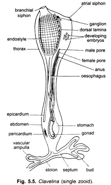
Clavelina (Fig. 5.5) is a colonial ascidian forming large clusters found on rocks in shallow seas of Europe. The colony consists of several independent individuals called zooids attached basally with the branching creeping stolon which is fixed to a rock. The zooids are free from each other, but all are connected by a common blood system. Each zooid is about 2.5 cm long and covered by a transparent test.
A zooid has an upper thorax and a lower abdomen connected by a middle narrow waist. The thorax has two short siphons, a pharynx with endostyle, dorsal lamina with languets and a circular dorsal tubercle. The abdomen is narrow and long and contains the rest of the alimentary canal, heart, gonad and stolon.
A narrow tubular outgrowth extends from the pharynx through the abdomen into the stolon, it is called epicardium. Gonad is hermaphrodite located in the intestinal loop. Reproduction is both sexual and asexual. The fertilised eggs develop in the atrial cavity, the tadpole larvae escape for a short free-swimming existence.
Thus, Clavelina is viviparous. The larva gets fixed and metamorphoses into a young oozoid. The oozoid forms a stolon. Within stolon are present two blood channels partitioned by a mesodermal septum. New zooids arise asexually from the terminal branches of the stolon to form a colony of blastozooids.
In autumn the zooids of a colony disappear, leaving the stolon into which the substance of zooids is passed as food-laden trophocytes which survive the winter. New zooids arise from this stolon next spring. Clavelina is of interest because it links the simple ascidians to compound ones.
Type # 6. Pyrosoma:
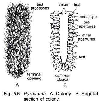
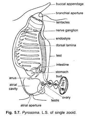
Pyrosoma (Figs. 5.6 and 5.7) (Gr., pyro = fire) is a pelagic, free swimming colony which emits bright, phosphorescent light when stimulated. The light is produced by symbiotic bacteria which inhabit the mesodermal cells and even invade the eggs. It is found in the warmer parts of Atlanic, Indian and Pacific represented by 2 species. The colony is tubular with only one end open having a velum. The colony floats horizontally.
The colonies range from 2.5 cm to 120 cm in length, they have an outer test. The test is gelatinous with many projecting processes on its outer surface. Embedded in the test or wall of the cylinder are numerous zooids called blastozooids, they are arranged in such a way that the branchial aperture of each opens outwards guarded by a tongue-like appendage and the atrial aperture into a common cloacal cavity of the cylinder which opens outside by a cloacal aperture regulated by a diaphragm or velum.
Colony locomotes by jet propulsion since a stream of water is shot out from the cloacal aperture. A blastozooid has a structure like an ascidian. It has a pharynx with many stigmata, an endostyle and a dorsal lamina with languets. At the inner end of the pharynx is an atrium. Heart lies behind the pharynx.
ADVERTISEMENTS:
Subneural gland and ocellus are also present. There are circum-oraland atrial muscles forming complete but feeble rings at the two ends. There is a hermaphrodite gonad with a single yolky ovum and a lobed testis. Near the endostyle is a stolon which forms additional blastozooids by budding.
Each blastozooid sieves water from the sea into the common cloacal cavity of the colony, then a stream of water is shot out from the open posterior end moving the colony forward by propulsion. Speed is increased by the velum narrowing the posterior opening in ejecting water.
In Pyrosoma there is no tailed larva, development is direct and takes place with in the body of the parent. The fertilised egg undergoes meroblastic cleavage and forms an oozooids or cyathozooid having a stolon. It has a primitive gut, a tubular nervous system and a pair of laterally placed atrial tubes. Within maternal body it feeds upon kalymnocytes (follicle cells). (A zooid arising from an ovum is an oozooid, while the one formed by budding is a blastozooid).
The stolon (anterior part of body) forms four buds called blastozooids, each of which has a stolon. The posterior part of the cyathozooids eventually atrophies. This colony of four individuals (tetrazooid colony) gets enclosed in a common test, it passes out through the cloaca of the parent colony. The cyathozooid begins to atrophy, but more blastozooids are produced by budding from the stolons of tetrazooid colony to form an adult Pyrosoma colony.
Type # 7. Doliolum:
Doliolum (Fig. 5.8) is pelagic, free-swimming and transparent creature. It is found in cold, tropical and subtropical seas.
Structure:
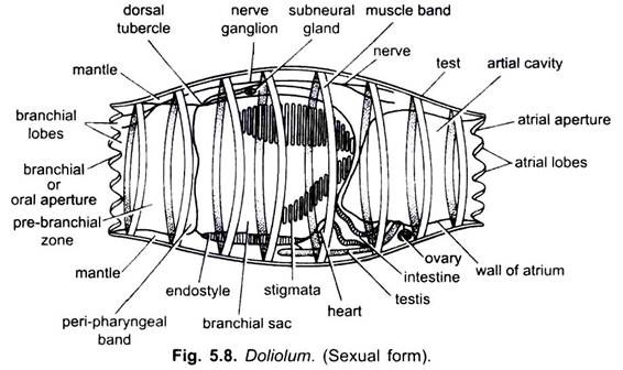
It is one to two cm long with a transparent barrel-shaped body covered over by a thin transparent test. The mouth and the atriopore are at the opposite ends. Both the apertures are surrounded by numerous lobes. Its life cycles include two phases- a solitary sexual gonozooid and a colonial asexual oozoid.
Gonozoid:
The sexual gonozooid or solitaria phase has a thin transparent test. The mantle or body wall has a single layer of epidermis and beneath it lies the mesenchyme which forms eight complete muscle bands, the first and last of which act as sphincters. Contractions of muscles drive water through the posterior end, thus, the animal moves by propulsion. The mouth and atrial apertures are at opposite ends of the barrel-shaped body, each has 10 or 12 sensory lobes.
Alimentary Canal:
Mouth leads into a pharynx having several elongated stigmata in its posterior wall, a ventral endostyle, but there is no dorsal lamina. Pharynx leads into oesophagus, ventrally it leads into a dilated stomach, tubular intestine which finally opens into the atrial cavity.
Nervous System:
There is a dorsal brain (cerebral ganglion) in the third or fourth inter-muscular zone. The brain has central fibrous medulla surrounded by the ganglionic cells forming cortex. The brain gives off nerves to the various organs. The branchial and atrial lobe has tactile sense organs.
Neural Gland:
It is present near the nerve ganglion and its duct opens into the anterior end of the branchial sac.
Heart:
The tubular heart is enclosed within a transparent, non-contractile tubular pericardium and lies at the posterior end of branchial sac. The heart gives off vessels from its both ends. Blood is colourless having clear plasma and a less number of amoebocytes.
Gonozooid is hermaphrodite. Gonads are found on the left side of the middle line close to the alimentary canal. Ovary is sac-like and the testis tubular. Gonoducts open into the atrium.
Reproduction:
It is specialised and highly complicated. The fertilised eggs of the sexual gonozooid develop in sea water. Cleavage is holoblostic resulting into blastula which undergoes aperture gastrulation by invagination. Later on gastrula develops into a tailed larva having a notochord. Its body is like that of the adult, i.e., barrel-shaped with nine muscle bands but there is a tail at the ventral hind end. Tail contains notochord formed of more than 10 chordal cells.
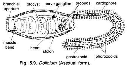
The body has two small processes, a postero-dorsal cadophore and a ventral stolon. In its pharynx only four gill-slits are present on each side. It possesses the statocyst on the lateral side. Oozooid (asexual phase) also possesses a dorsal appendix (cadophore) in between seventh and eighth muscle bands.
It is immovable elongated structure and encloses a part of coelom. Stolon is present on the ventral side. The larva after a little metamorphosis loses its tail but does not get fixed, it changes into an asexual oozooid stage or gregaria phase which acts as a float and reproduces asexually.
Asexual Reproduction by Budding:
In the asexual oozooid the cadophore and stolon increase. The stigmata, endostyle and alimentary canal are lost. There are nine complete muscle bands which increase in thickness, and nervous system attains a higher development. Stolon’s free end undergoes segmentation to form small constricted bodies which get detached from the stolon as probuds.
The probuds migrate along the body and get fixed to the cadophore. The probuds divide several times and get fixed as lateral and median rows of buds on the cadophore. These buds are of three kinds. The lateral buds develop into small Doliolum-like zooids with rudiments of gonads that soon atrophy.
Alimentary canal develops. These are called trophozooids or gastrozooids and these are helpful in the nutrition and respiration of the colony. Some of the median zooids called phorozooids act as nurses and become detached.
These are barrel- shaped in structure with 8 muscle bands and vascular structures. They have no gonads. The other median zooids called gonozooids are fixed on the stalks of phorozooids and carried for a time. The gonozooids acquire gonads, separate from phorozooids and form the adult sexual stage or solitaria phase.
Doliolum exhibits polymorphism and alternation of generations in its complicated life history. The sexual Doliolum phase reproduces sexually and develops asexual phase which is polymorphic- nutritive gastrozooids, foster phorozooids and sexual gonozooids.
Type # 8. Salpa:
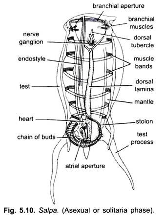
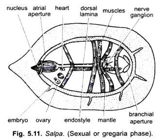
Salpa (Fig. 5.10 and 5.11) is found in almost all seas as a free-swimming pelagic animal. It occurs in two phases- an asexual oozooid or solitaria form and a sexual blastozooid or gregaria form.
Structure of Asexual Salpa:
Salpa is a transparent, small pelagic marine creature. Its body is covered by a soft gelatinous test closely attached with the mantle. The body is ovoidal having branchial and atrial apertures at the opposite ends. The nucleus is opaque which contains the heart and the alimentary canal.
Muscles bands are six to nine C-shaped, incomplete ventrally. Atrial band is complete, while the oral band is prolonged into lips and acts as sphincter. Their contractions eject water through the atrial aperture and the animal is propelled forwards.
Digestive System:
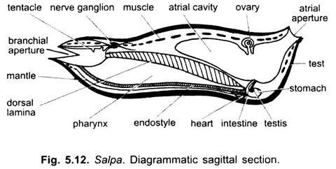
Branchial aperture is wide and guarded by movable upper and lower lips. It leads into a tubular buccal cavity which is followed by prebranchial zone of pharynx and then large elongated pharynx. It has a dorsal lamina and a central endostyle. The side walls of pharynx have no stigmata and freely communicate with the atrial cavity on each side.
Dorsal tubercle is located near the tentacular languet formed by the anterior end of dorsal lamina. Beneath it lies two sub-neural glands which open independently into the pharynx. Above these is present the nerve ganglion. Rest of the alimentary canal includes the oesophagus, saccular stomach having two lateral caeca, looped intestine and rectum opening into atrium through anus.
Nervous System:
Nerve ganglion lies near the anterior end gill and gives off many nerves. Above the nerve ganglion is present a U-shaped pigmented ridge-like eye. In some species one or more lateral ocelli are also present.
Heart:
It is present near the stomach. It gives off many vessels to the various organs of the animal. Branchial sac receives many transverse vessels from the dorsal and central aorta.
Reproduction:
The asexual oozooid has no gonads, but it has a stolon arising from near the endostyle. The stolon elongates and segments into a chain of buds, which break off in groups to form the sexual blastozooids or gregaria stage which later separate.
Sexual Form:
The sexual blastozooid has a testis and an ovary with a single ovum, but it has no stolon. Ovary is unpaired, located near the right side of nucleus. It opens through oviduct into the atrium. Testis is a paired tubular branched structure near the nucleus, on either side of intestine.
Both open independently into the atrium. Sexual blastozooid has a general structure like an asexual oozoid, but it differs in being smaller. The body is asymmetrical with no test processes, number of muscle bands is fewer, gonads are present and there is no stolon. It produces a single egg at a time which is cross fertilised internally because the animal is protogynous (i.e., ova matures first).
Fertilisation:
Fertilisation is internal. Before fertilisation the stalk-like oviduct shortens and draws the ovary towards the dorsal side of the nucleus. The ovary is now enclosed in a lumen having a relation with the atrium. Fertilisation of a single ovum occurs within this lumen. After fertilisation the oviducal opening becomes closed and ovarian epithelium forms a sac-like structure around the ovum.
Development:
Development is direct and takes place within the body of the parent. Cleavage is holoblastic. The developing embryo projects into the branchial cavity. The nourishment of embryo occurs by the formation of a diffusion placenta through which a close union is brought about between the vascular system of the parent and that of the embryo. The placenta of Salpa is partly formed from follicle cells and ectoderm cells of the embryo, and partly from the cells of oviduct.
Segmentation is complete. During segmentation there is a migration inwards of some of the cells of the follicle and of the wall of the oviduct. These cells enter the segmenting ovum and pass among the blastomeres. These cells are called kalymnocytes and possibly they carry nourishment to the developing embryo. These are broken up and eventually completely absorbed.
There is no tailed larval stage. The embryo develops the muscle bands and all the characteristic parts of the adult while present within the parents body. The sexually produced embryo grows into a solitary asexual oozoid which escapes from the parent and becomes free-swimming. After a time there grows a stolon, on the surface of which are formed a number of bud-like projections. These increase in size as the stolon elongates and each takes the form of a sexual Salpa. The chain of zooids formed on stolon breaks off in groups and swim as such while reproductive organs develop in each.
Salpa is, thus, dimorphic and it exhibits an alternation of generations in its life history.
Type # 9. Oikopleura:
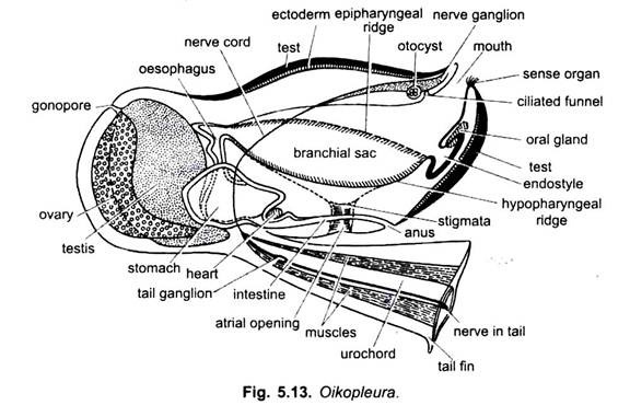
Oikopleura (Fig. 5.13) is pelagic free-swimming and neotenous found in almost all seas except Polar Regions.
House:
The house is formed of a gelatinous transparent test which is cuticular and not made of tunicin.
Body:
The animal is about 2 mm long and surrounded by a large house (loose test) which does not adhere to the animal. The large “house” has a pair of funnel-shaped incurrent apertures having a sieve of fine threads which allows only minute particles to enter along with water. Movements of the tail cause food and water to enter the house through the incurrent apertures.
Inside the house is an elaborate filtering apparatus formed of sieve tubes which filters minute organisms which are sucked into the mouth due to ciliary action of stigmata. Food particles are entangled in mucus produced by the endostyle, the food-laden mucus cords are drawn into the stomach. When pressure of water increases in the house, a door opens at the tail end and water passes out, this jet of water also propels the house forwards.
The house also has an emergency exit door through which the animal can escape, it never re-enters the house but its ectoderm secretes a new house with great rapidity. The house is protective, hydrostatic and for filtering food. The animal has an oval trunk and a laterally compressed tail attached to the ventral side, near the hind end. An anterior mouth leads into a large pharynx having a short endostyle, two peripharyngeal bands, and two ventral ciliated stigmata which open directly to the exterior because there is no atrium.
There is no dorsal lamina. Rest of the gut has an oesophagus, stomach and intestine opening to the exterior by an anus in front of the stigmata. A cerebral ganglion lies near the mouth; close to it is a statocyst. Neural gland is absent. Heart lies beneath the stomach.
At the posterior side of the body is a large hermaphrodite gonad, and the animal is protandrous. (Oikopleura dioica is not hermaphrodite; it has separate males and females). The vibratile tail is laterally compressed and twisted. It is surrounded by a wide tail fin. The tail is supported by notochord and also contains a ganglionated nerve cord and unsegmented muscles. The larva does not undergo metamorphosis since it is neotenous. It becomes sexually mature due to the formation of gonad.
