In this article we will discuss about:- 1. External Features of Ascidia 2. Body Wall and Atrial Cavity of Ascidia 3. Digestive and Respiratory System 4. Respiration 5. Blood Vascular System 6. Excretory System 7. Nervous System 8. Reproductive System 9. Metamorphosis.
Contents:
- External Features of Ascidia
- Body Wall and Atrial Cavity of Ascidia
- Digestive and Respiratory System of Ascidia
- Respiration in Ascidia
- Blood Vascular System in Ascidia
- Excretory System of Ascidia
- Nervous System of Ascidia
- Reproductive System of Ascidia
- Metamorphosis of Ascidia
1. External Features of Ascidia:
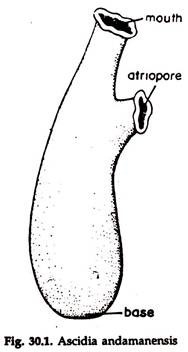
Body bag-like, with a basal end attached to some support and the free end directed upwards (Fig. 30.1). Two apertures are present; an eight-lobed oral funnel or oral siphon at the top and a six-lobed atrial funnel or atrial siphon placed laterally at some distance from the free end, towards the base.
ADVERTISEMENTS:
The atripore is present in the middle of the atrial funnel. The body is enclosed in a tough but translucent tunic or test. Water enters the body through oral funnel and goes out through atrial funnel.
2. Body Wall and Atrial Cavity of Ascidia:
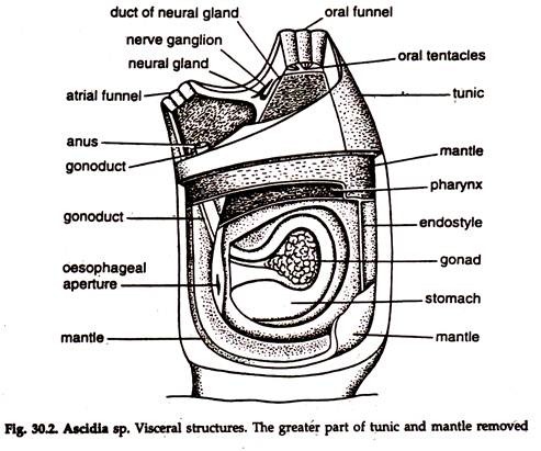
The body wall is composed of an outer tunic and an inner mantle (Fig. 30.2). The tunic is secreted by epidermis, made of tunicin (cellulose and glycoprotein) and contains scattered cells. The cells in the tunic are mesodermal in origin and migrate through the ectoderm to the outer surface.
ADVERTISEMENTS:
Outgrowths of the body wall with prolongations of blood channel enter the test and ramify in all direction. A longitudinal septum divides a channel into two and each ends in a small bulb. Blood moves-along one passage, circulates in the bulb and returns through the second passage.
The soft body wall or mantle underlies the test. It remains suspended in the test, being firmly attached to it only round the oral and atrial funnels. Ectoderm cells with underlying layers of connective tissue containing muscle fibres constitute the mantle. The mantle is produced into two short but wide tubular projections—the oral and atrial funnel.
The prolongations are continuous with the margins of the apertures of the test and guarded by strong sphincter muscles, capable of closing the openings. The mantle encloses an atrial or peribranchial cavity, atrium, opening to the exterior through atriopore. The atrial cavity is formed by the involution from the outer surface.
ADVERTISEMENTS:
Coelom:
The coelom is greatly reduced and restricted to pericardial and gonadal cavities.
3. Digestive and Respiratory System of Ascidia:
Mouth, stomodaeum, pharynx, oesophagus, stomach, intestine and anus constitute digestive system:
Mouth and stomodaeum:
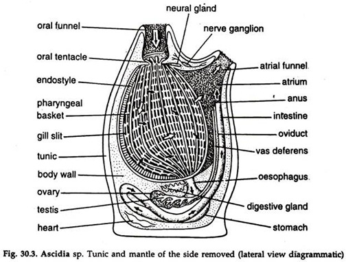
The mouth is surrounded by velum, located in the posterior end of the oral funnel. The velum bears velar tentacles. Sphincter muscles regulate opening and closure of the mouth. The mouth leads to a short but wide oral passage, the stomodaeum, which communicates with a large chamber, the pharynx or branchial chamber (Fig.30.3).
Pharynx:
The pharynx extends posteriorly near to the base of the body and remains attached to the mantle along the ventral side. The space between the mantle and pharynx is called atrium. Vascular mesodermal tissue, the connectives or trabeculae run from the body wall to the wall of the pharynx across the atrium. The wall of the pharynx bears numerous slit-like vertically disposed apertures, the stigmata, arranged in transverse rows.
ADVERTISEMENTS:
The edges of the stigmata bear numerous strong cilia. By the end of metamorphosis, two primary gills, protostigmata appear on each side of the pharynx. The number increases to six on each side, by the development of independent perforations.
With growth, the six protostigmata elongate and subdivide to form a row of definitive stigmata; additional rows continue to form as growth proceeds, and the perforated pharynx is produced.
Along the line of adhesion of the pharynx with the mantle, a thickening in the form of a pair of ventral longitudinal folds, separated by a groove, are present on the inner surface of the pharynx. The grooved thickening is called endo-style. Two rows of mucous cells separated by a row of ciliated cells are present on each side of the endo-style.
In the median groove of the endo-style, a group of cells with long cilia are present. A ciliated fold, dorsal lamina is present on the inner surface of the pharynx along the mid-dorsal line. Pharynx acts as both food capturing and respiratory organ. It leads to oesophagus.
Oesophagus:
The oesophagus is short and narrow and located near the posterior end of dorsal lamina. The oesophagus joins the stomach. The oesophagus, stomach and intestine are embedded in the mantle on the left side.
Stomach:
The stomach is large, fusiform, with a thick glandular wall.
Intestine:
The intestine is narrow, thin-walled, bent around in a double loop and runs forward to open in anus in the atrial cavity. A thickening along the inner wall of the intestine forms the typhlosole. Delicate ramified tubules, the pyloric glands over the wall of the intestine, open into the stomach through a duct.
Feeding in Ascidia:
Beating movement of cilia in the stigmata create a water current, the respiratory and food current, which enters the pharynx through mouth and stomodaeum and from there goes to the atrium through stigmata, and thence to the exterior via atripore and atrial funnel.
The gland cells of the endo-style secrete viscid mucus. Active, lateral beating movement of the cilia, distributes the secretion on the side wall of the pharynx; the median ventral cilia assist to deflect the mucus to either side of the groove.
On reaching the wall of the pharynx, the mucus is driven upwards and towards the mid-dorsal line as a sheet. Additional mucus follows the trail. Food particles carried by water current are entangled in the mucus sheet. The dorsal lamina along the mid-dorsal line of the pharynx collect the food-laden mucus and conducts it backward, towards the oesophagus.
Digestion and Absorption in Ascidia:
Digestion occurs in stomach. Strong carbohydrates and weak protease and lipase are secreted by two glands in the stomach. The pyloric gland is possibly both digestive and excretory in function. Absorption takes place in intestine.
4. Respiration in Ascidia:
The walls of the stigmata are highly vascularized. Dissolved oxygen in the inward water current is absorbed in the stigmata and carbon dioxide is released, which is moved out by the outward current.
5. Blood Vascular System of Ascidia:
The blood vascular system is well-developed and consists of a heart, blood vessels and sinuses:
Heart:
The heart is a fusiform muscular sac in the pericardium and located near the stomach. A large vessel arises from each end of the heart. The branchio-cardial vessel arising from the ventral portion of the heart runs along the mid-ventral line of the pharynx, below the endo-style and gives off branches running along the bars between the rows of stigmata. Smaller branches arising from these branches run between the stigmata of each row.
The cardio-visceral vessel from the dorsal end of the heart breaks up into branches and ramify on the alimentary canal and other organs. The vessels or lacunae open into a large sinus, the viscero-branchial sinus. The sinus rims along the mid-dorsal wall of the pharynx and communicates with the dorsal ends of the series of transverse branchial vessels. In addition to the principal vessels, numerous lacunae extend throughout the body including the test.
The contractions of the heart are peristaltic. The contraction follows from one end of the heart to the other end for a certain period, which is followed by a pause. The next contraction takes place in opposite direction. In this way, the course of blood in the heart is reversed at regular intervals.
Blood:
The plasma is colourless and the corpuscles are few. Lymphocytes, phagocytes, macrophages, vacuolated compartmental cells and coloured and colourless cells are present.
6. Excretory System of Ascidia:
Nephrocytes present in blood are concerned with excretion. Particles of urates and xanthine in the cells-are disposed of by storage in the form of concretions in special excretory vesicles or renal organs.
7. Nervous System of Ascidia:
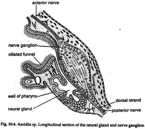
The nervous system is extremely simple. A neural ganglion (Fig. 30.4) lies between the oral and atrial funnels, embedded in the mantle. It is dorsoventrally elongated and gives off nerves at each end, which innervate various parts of the body.
Neural gland:
The neural gland lies on the ventral side of the neural ganglion and considered homologous with at least a part of the hypophysis of vertebrates by some workers, which is controversial. A duct runs forward from the neural gland and opens into the cavity of the pharynx through a ciliated funnel. The duct is folded upon itself to form a prominent dorsal tubercle projecting into the pharyngeal cavity.
8. Reproductive System of Ascidia:
The sexes are united. The ovary and testis are closely situated on the left side of the body in the intestinal loop (Fig. 30.2, 30.3). The gonoduct—oviduct or spermiduct is long, continuous with the gonad and opens in the atrial cavity close to the anus. Self-fertilization is prevented by production of ova and sperms at different times. Fertilization is external. Self- fertilization has been reported in some species of Ascidia.
Fertilization and Development:
Small, yolkless eggs float on water and in the majority, fertilization takes place there.
Development is indirect and associated with metamorphosis.
Segmentation is holoblastic and nearly equal. Only a few cells form the blastula. Gastrulation is by invagination. The gastrula elongates and, about three days after fertilization, a fish-like tadpole larva hatches out.
Larval form:
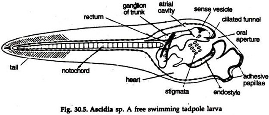
The tadpole larva (Fig. 30.5) is highly motile and moves with the help of tail. It does not take food for some time. The stage is known as non-feeding form.
1. The body is covered by tunic and divisible into an oval head and a Jong tail.
2. Three adhesive papillae or chin warts; one mid-dorsal and two ventrolateral are present in front of the head.
3. The tail is laterally compressed, pointed terminally and provided with a caudal fin continuous with dorsal and ventral fins.
4. Striae—presumably precursors of fin rays of fishes—are present in the firis.
5. The central nervous system consists of an enlarged, anterior sense vesicle opening in the pharynx by neuropore and a narrow, hollow caudal part (spinal cord).
6. A median eye with retina, cornea and lens and a statocyst, organ of balance are present in the sense vesicle.
7. An endodermal notochord is restricted in the tail region, extending up to the pharynx and en-sheathed in a gelatinous matter.
8. Segmental muscles arranged on both sides of the nerve cord are present in the tail region.
9. The alimentary canal is divisible into two regions. The anterior region consists of a mouth and a well-developed pharynx with two pairs of gill slits and an endo-style, and the posterior region gives rise to oesophagus, stomach and intestine.
10. Atrial sacs are paired.
The tadpole larva is positively phototactic and negatively geotactic. After a brief active period, the larva becomes sluggish and Marts searching for a suitable substratum for fixing itself. The preference is always for hard or rocky one. The larva fixes itself to the support with adhesive papillae, turns negatively phototactic and positively geotactic and metamorphosis begins.
9. Metamorphosis of Ascidia:
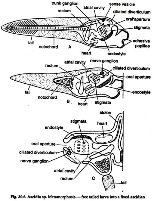
Metamorphosis (Fig. 30.6) of tadpole larva in Ascidia is a two-way process. As the majority of the organs of the larva are lost, the process has been termed as retrogressive. But at the same time some of the structures further develop, which may be considered as progressive.
Changes in the larva following fixation:
Retrogressive metamorphosis:
1. The length of the tail is considerably shortened, the number of striae reduced and finally the tail disappears.
2. The notochord becomes coiled, disorganized and disappears with the tail.
3. The nerve cord becomes restricted to the trunk region and gradually reduced to a solid ganglion.
4. 90° shifting of the mouth occurs from the point of attachment due to rapid growth of the region between adhesive papillae and mouth and also suppression of growth of the original dorsal side.
Progressive metamorphosis:
1. The pharynx enlarges in size and the number of stigmata increases.
2. Atrium enlarges.
3. Velum appears.
4. Neural gland differentiates.
5. Reproductive organs gradually develop.
In course of metamorphosis the active, free-swimming larva with a notochord, a well-developed nervous system and complex organs of special senses is degenerated to a fixed, non-motile adult, in which all the chordate characters disappear, except gill slits, endo-style and structures associated with feeding.
