In this article we will discuss about:- 1. Description of Lamprey 2. External Features of Lamprey 3. Body Wall and Muscles 4. Digestive System 5. Respiratory System 6. Blood Vascular System 7. Excretory System 8. Nervous System 9. Reproductive System 10. Metamorphosis.
Contents:
- Description of Lamprey
- External Features of Lamprey
- Body Wall and Muscles of Lamprey
- Digestive System of Lamprey
- Respiratory System of Lamprey
- Blood Vascular System of Lamprey
- Excretory System of Lamprey
- Nervous System of Lamprey
- Reproductive System of Lamprey
- Metamorphosis in Lamprey
1. Description of Lamprey:
Lamprey, also called lamper eels belongs to family Petromyzontidae, order Petromyzontia, class Cyclostomata, superclass Agnatha, subphylum Craniata (Vertebrata), phylum Chordata. They are most common in the European river system.
ADVERTISEMENTS:
The marine form of sea-lamprey (P. marinus) is about one metre long; the fresh water lamprey (Lampetm Jluviatilis) is about 90 cm long and L. planeri, a lesser fresh water form is about 45 cm in length.
Two distinct phases are present in the life-history of lampreys. They spend major part of their adult life in sea and are provided with a suctorial mouth. The larval phase, the Ammocoetes lives in fresh water and feed on microscopic food trapped in endostyle.
2. External Features of Lamprey:
The head and trunk are nearly cylindrical and the tail is laterally compressed (Fig. 32.1). The smooth, slimy body surface is devoid of scales. The upper surface, is usually dark and the lower surface white. A median dorsal fin divided into two unequal parts and a caudal fin continuous with posterior dorsal fin are present. The fins are supported by fine cartilaginous fin rays.
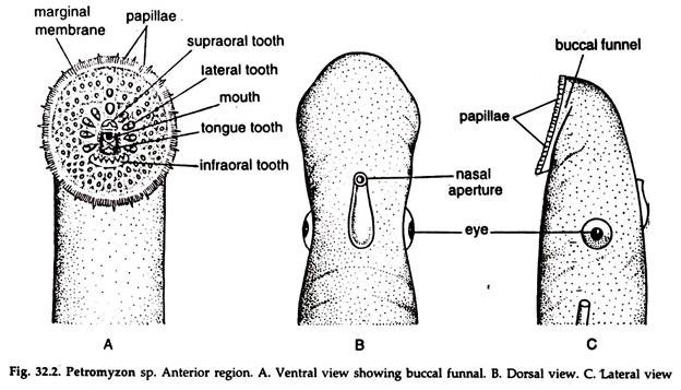
A single median nostril and the pineal organ; closely behind it are present on the dorsal surface of the head. Eyes are paired, lateral, without eyelids but covered with a’ transparent skin (Fig. 32.2B, C).
At the anterior end a large downwardly directed basin-like depression, the buccal funnel is present (Fig. 32.2A). It is surrounded by a marginal membrane, overlapping the oral fimbrae and sensory cirri arising from outside the marginal membrane.
Radiating rows of yellow, homy teeth on cartilaginous pads are present in the buccal funnel. A prominence, the tongue bearing large, horny teeth projects from the bottom of the buccal funnel. The narrow mouth lies just above the tongue.
ADVERTISEMENTS:
Gill slits or gill pores are seven pairs of small openings on the sides of the head.
The anus, a narrow opening lies in a small depression, the vent on the ventral surface at the junction of trunk and tail.
A small papilla bearing urinogenital aperture lies immediately behind the anus.
Segmental sense organs, the lateral line sense organs on either side of the body and head are exposed to the exterior, not enclosed in lateral line canals.
3. Body Wall and Muscles of Lamprey:
The skin is soft. The epidermis bears numerous mucus secreting unicellular glands.
In addition to these, two other types of secretory cells are present:
(a) Granular cells of unknown function and
(b) Elongated club cells with hyaline cytoplasm, probably secreting slime.
ADVERTISEMENTS:
A layer of collagen fibres form the cutis beneath the epidermis. Pigment cells or chromatophores are present in this layer. The pigment cells are star-shaped and change their position under different conditions of illumination, imparting change of colour from pallid to dusky and vice versa.
The muscles of the trunk and the tail are arranged in myotomes in a zigzag course.
Locomotion:
The lamprey swims by producing waves of curvature from anterior to posterior of the body, alternately on each side, due to contraction of myotomal longitudinal muscle fibres. Since the waves are short, progress by swimming is slow.
Endoskeleton:
The skeleton is wholly cartilaginous. The notochord, ill-developed skull and branchial basket constitute axial skeleton.
Notochord:
The notochord (Fig; 32.4) is persistent, un-segmented and enclosed in a tough sheath of an inner fibrous and an outer elastic layer. Small, segmentally arranged, vertical cartilaginous rods are attached to the sides of the notochord bounding the spinal canal. In the caudal region the rods fuse to form a single plate bearing foramina for the spinal nerves and sends processes to the base of the fin.
Cranium:
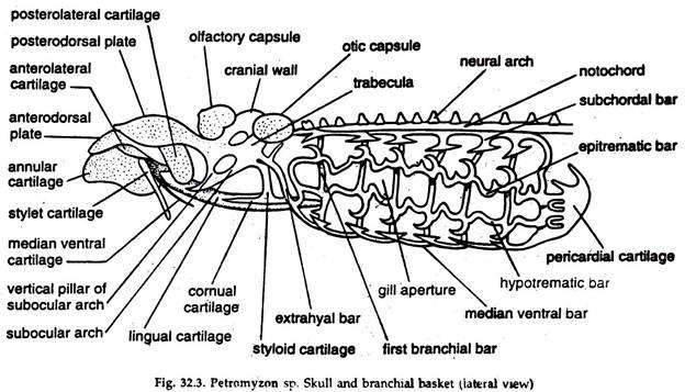
The cranium is primitive. The floor is occupied by a basal plate formed by paired parachordals and trabeculae. An incomplete box enclosing the brain and special sense organs is formed by pieces of cartilage attached to the basal plate (Fig. 32.3).
A large aperture, die basicranial fontanelle is present in front of the cranium. The roof of the cranium is made of membranous fibrocartilage, supported by a transverse bar. Auditory capsules are united with the posterior end of die basal plate and form the end of the neurocranium.
The olfactory capsule is an imperfectly paired concavo- convex plate, supporting the posterior wall of the olfactory sac. Fibrous tissue joins the capsule with the cranium.
A sub-ocular arch extends downward and outward from each side of the nasal plate to support the eye (Fig. 32.3). A slender styloid process hangs from the sub ocular arch and a small cornual cartilage is connected to its lower end. The skeleton of the buccal funnel and branchial basket are attached to the cranium.
A ring-like annular cartilage supports the buccal funnel. The tongue is supported by a long, impaired lingual cartilage.
Visceral skeleton:
The visceral skeleton (Fig. 32.3) consists of a branchial basket of nine irregular, vertical cartilaginous bars or rods on each side. The first rod is located immediately posterior to the styloid process, the second in front of the first gill slit and the rest posterior to the remaining gill slits.
The vertical bars are joined by longitudinal bars. Posteriorly the branchial basket extends to form a cup-like pericardial cartilage accommodating the heart.
Coelom:
The coelom is represented in two cavities, a small, anterior pericardial cavity containing heart and a large posterior pleuroperitoneal cavity containing visceral organs.
4. Digestive System of Lamprey:
An alimentary canal, a pair of buccal glands, a liver and patches of secretory cells constitute the digestive system:
Alimentary canal:
It is a nearly straight tube starting at mouth and ending in anus.
Buccal funnel:
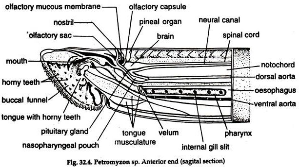
A large, downwardly directed basin-like depression (Fig. 32.4) at the anterior end of the body. The mouth is a narrow opening above the tongue, projecting from the bottom of the buccal funnel.
Radiating rows of horny, yellow teeth on cartilaginous pad are present in the buccal funnel. The teeth, when worn out, are succeeded by others developing at their bases. The tongue also bears large, horny teeth.
Buccal cavity, oesophagus and respiratory tube:
The mouth leads into a buccal cavity, communicating behind with two tubes, placed one above the other (Fig. 32.4). The dorsal one is the oesophagus and the ventral one, respiratory tube, which is closed behind. The opening of the respiratory tube is guarded by a curtain-like velum bearing velar tentacles. The oesophagus leads into an un-convoluted intestine through a valvular aperture.
Intestine:
The intestine is slightly dilated anteriorly. It bears a typhlosole, which takes a slightly spiral course lengthwise and known as spiral valve. The intestine widens posteriorly to form the rectum ending in anus, in a small depression, the vent.
Digestive glands:
Paired buccal or salivary glands, a voluminous liver and patches of secretory cells in the epithelium of the anterior part of intestine constitute digestive glands.
Salivary or buccal glands:
They are paired and embedded in the hypobranchial muscles and open separately by ducts in the buccal funnel, below the tongue. The walls of the glands are folded and the secretion containing anticoagulant, presumably prevent coagulation of blood sucked from the host.
Liver:
The liver is voluminous, bilobed and lacks bile duct in the adult, though the ducts are present in the larva.
Pancreas:
Definite pancreas is wanting but patches of cells comparable to pancreatic acini are present in the epithelium of the anterior part of the intestine.
Feeding and digestion:
The Petromyzon has a suctorial mouth, with a rasping apparatus. It clings to fishes and feeds on their tissues. The digestive enzymes secreted by the liver and proteolytic enzymes produced by zymogen granules in the so-called pancreatic acini help in digestion of carbohydrates and proteins.
5. Respiratory System of Lamprey:
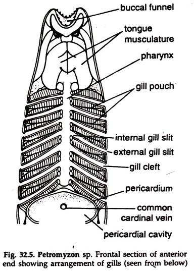
The respiratory system consists of seven pairs of gill pouches or branchial pouches (Fig. 32.5). In the adult connection between the respiratory tube and oesophagus is lost.
A gill pouch has the shape of a biconvex lens with numerous gill lamellae on the’ inner surface. The gill pouches are separated from one another by inter-branchial septa. Each gill pouch opens to the exterior separately through gill pores or gill slits.
Mechanism of Respiration:
Both incurrent and ex-current streams of water pass through gill pouches. The branchial skeleton is elastic. With the expansion of the branchial basket water enters the respiratory tube through gill pores. The water is expelled through the same route by contraction of the branchial basket. Gaseous exchange takes place in the gill lamellae.
6. Blood Vascular System of Lamprey:
The circulatory system is much advanced and consists of a heart, arterial and venous systems:
1. Heart:
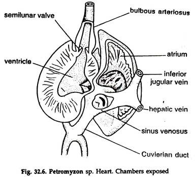
The heart is two-chambered (Fig. 32.6), enclosed in a pericardium and situated posterior to the last pair of gill pouches. The chambers are an atrium and a ventricle. The atrium lies left of the ventricle and receives blood from a small, thin-walled sinus venosus. The atrium opens into the thick-walled ventricle. The slightly dilated base of the ventral aorta arising from the ventricle is bulbous arteriosus.
The slit-like sinuatrial aperture is guarded by a pair of sinuatrial valves. The small atrioventricular aperture is guarded by atrioventricular valves. A single set of semilunar valves at the base of the bulbous prevents backflow of blood to the ventricle.
Blood:
The colour of blood is red due to the presence of haemoglobin in erythrocytes, which is intermediate between the haemoglobin of invertebrates and gnathostomata. The red blood corpuscles are nucleated and circular. The white blood corpuscles are almost like the lymphocytes and polymorphs of higher vertebrates. The blood cells are formed in the tissues present in spiral valve, kidneys and spinal cord.
2. Arterial system:
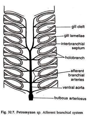
The ventral aorta with a slightly dilated base, the bulbous arteriosus arises from the ventricle between the last pair of gill pouches. Eight pairs of afferent branchial arteries are given off from the ventral aorta, ending in capillaries in the gills.
Afferent branchial arteries:
The ventral aorta leaves the heart as a single vessel, which bifurcates at the level of the fourth gill pouch (Fig. 32.7). From each of the paired anterior branch arise four afferent branchial arteries and four pairs are given off from the unpaired portion.
The first of the eight vessels on each side sup plies the gill lamellae of the anterior hemi branch of the first gill pouch, while the last ends in most posterior hemi branch. Each of the six remaining afferent branchial arteries divides and supply the lamellae on either side of the septum, i.e. the posterior hemi branch of a gill pouch and anterior hemi branch of the next pouch.
Efferent branchial arteries:
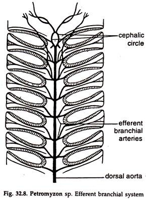
These vessels collect blood from the gill lamellae and join to form a single median dorsal aorta (Fig. 32.8). The anterior end of the dorsal aorta is paired for a short distance and again join to form the cephalic circle. Arteries supplying brain, eyes, tongue and parts of the head arise from the cephalic circle. The dorsal aorta gives off a series of segmental arteries to the myotomes and sends branches to gut, kidneys and gonads.
3. Venous system:
The venous system consists of true veins and network of venous sinuses. The caudal vein bringing back blood from the tail region and running anteriorly divides into two posterior cardinal veins in the posterior end of the abdominal cavity. The cardinals receive blood from myotomes, kidneys and gonads and open into the heart by a single ductus Cuvieri on the right side (Fig. 32.6). The left ductus Cuvieri is absent in adult.
A pair of anterior cardinal veins (jugular) drain blood from the anterior region of the body. A large median inferior jugular vein brings back blood from the musculature of the buccal funnel and gill pouches. Renal portal system is absent. A hepatic portal vein drains blood from the gut and receives a vein from the head.
A contractile portal heart is present in the hepatic portal vein. A single hepatic vein collects blood from the liver and joins the sinus venosus.
The sinus venosus receives three vessels:
(a) A large single common cardinal vein on the dorsal side,
(b) A small inferior jugular vein on its anteroventral side and
(c) A small hepatic vein.
Only deoxygenated blood passes through the heart. Blood received from different parts of the body by sinus venosus is sent to atrium ventricle → ventral aorta → gills → body. It is a single type circulation.
7. Excretory System of Lamprey:
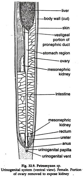
The excretory system consists of a pair of kidneys and their ducts. The kidneys are mesonephric in origin. They are long, strap-shaped and lie on either side of the mid-dorsal line (Fig. 32.9), from which they are suspended by mesentery-like membrane.
The renal units of adult kidney are renal corpuscles. A mesonephric duct, the ureter, runs along the free edge of each kidney. In the adult, vestiges of pro- nephric ducts of larva are present anterior to the functional part, or mesonephric kidney.
The two ureters open into urinogenital sinus, a cavity within the uringenital papilla, just behind the rectum. The urinogenital papilla opens to the exterior by a urinogenital aperture, behind the anus in the vent. The urinogenital sinus is in communication with the coelom by genital pores, one on each side.
8. Nervous System of Lamprey:
The nervous system is well-developed and similar to that found in the vertebrate series though some of the structures are in a relatively primitive condition.
The central nervous system and peripheral nervous system constitute the nervous system:
1. Central nervous system:
It consists of a brain and a spinal cord enclosed in a single membrane, the menix. The nerves are non-myelinated.
Brain:
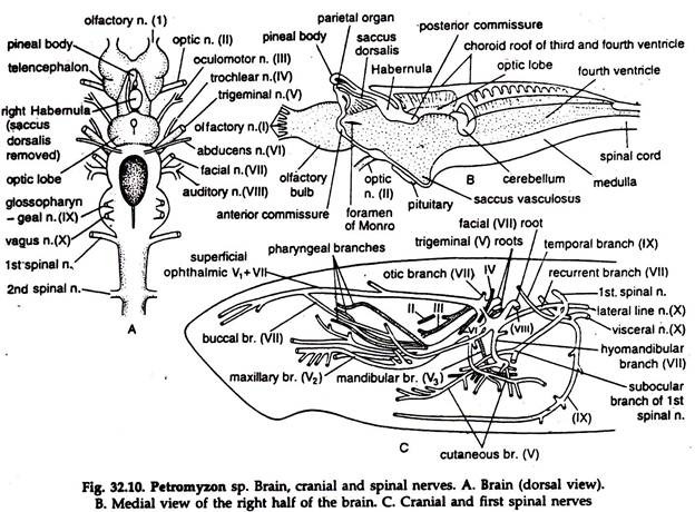
The brain (Fig. 32.10A, B) is divided into three distinct parts.
a. Forebrain (anterior portion) or pros encephalon.
b. Midbrain (middle portion) or mesencephalon.
c. Hindbrain (posterior portion) or rhomb encephalon.
Pros encephalon:
It consists of an anterior telencephalon made of two cerebral hemispheres, the two together is known as cerebrum, and a posterior diencephalon. The cerebral hemispheres are large and lobed. A large olfactory bulb, organ of smell is attached to each cerebral hemisphere anterolaterally.
The diencephalon has a membranous roof, projecting outwards as saccus dorsalis. The lateral sides are known as thalamus and the ventral part is formed by a well-developed hypothalamus bearing inconspicuous inferior lobes. The hypophysis or pituitary gland is closely applied to the hypothalamus. Two ganglia Habernulae, the right being much larger than the left are present on the dorsal border of the lateral walls.
These constitute epithalamus and are connected to the pineal apparatus. The pineal apparatus consists of a pineal eye lying against the transparent area of the cranial roof and the smaller Para pineal body, below and behind it. A simple crossing of optic nerves, not forming a chiasma, is present on the ventral surface.
Mesencephalon:
The midbrain bears two imperfectly divided optic lobes. A membrane, choroid plexus lies between the optic lobes. It is fused with the plexus over the fourth ventricle, the cavity of the medulla oblongata. Mesencephalon is concerned with sight.
Rhomb encephalon:
It is divisible into an anterior metencephalon (cerebellum) and a posterior myelencephalon (medulla oblongata). The cerebellum is small and occurs as a transverse band roofing over the anterior end of the fourth ventricle. The medulla oblongata is largest and highly developed. It controls the powerful sucking structures through trigeminal nerve.
Cavities of the brain:
The cavities of the brain are ventricles and that of the spinal cord is central canal.
Spinal cord:
The spinal cord is tubular, of fairly uniform diameter throughout its length, tapering gradually towards the posterior, and encloses the central canal. It is flattened, convex dorsally and concave ventrally. Grey matter is in the form of a longitudinal band surrounded by white matter, the two not sharply differentiated.
2. Peripheral nervous system:
Cranial nerves and spinal nerves constitute peripheral nervous system.
Cranial nerves:
Cranial nerves (Fig. 32.10C) are ten pairs as in some other vertebrates. Optic chiasma absent; trigeminal and facial nerves are intimately associated and the roots of the branches are not distinguishable. Vagus originates as a single nerve, branching subsequently.
Spinal nerves:
The dorsal and ventral roots of spinal nerves alternate with each, other, the dorsal root being somewhat posterior to the ventral root and they do not join. The dorsal root bears a ganglion.
In the gill region, fibres from adjacent dorsal roots unite to form a nerve (sensory). A nerve (motor) is formed by the union of fibres of adjacent ventral roots. Thus two nerves, one sensory and one motor are formed. These together form the hypobranchial nerve, innervating ventral part of the gill region.
Autonomic nervous system:
Segmental sympathetic ganglia are present. They are not connected with one another and sympathetic trunks are lacking. Preganglionic sympathetic fibres emerge from the spinal cord through dorsal spinal nerves.
Receptors and Sense Organs in Lamprey:
Olfactory sac (chemoreceptor), eyes (photoreceptors), pineal eye (photoreceptor), ears (equilibrium and phonoreceptor), lateral line sense organs (rheoreceptors), integumentary photoreceptors and gustatory receptors constitute receptor organs:
Olfactory sac (Nasopharyngeal sac):
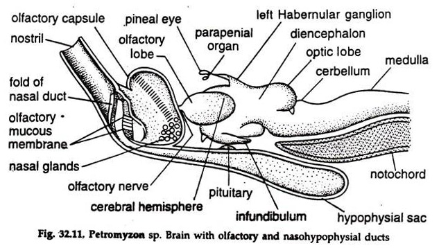
The external nostril communicates with a round nasal or olfactory sac by a short passage. Unlike other vertebrates, the lumen of the hypophysial rudiment, which develops from evagination of buccal epithelium, is continuous with the nasal sac, through a duct, the nasopharyngeal canal (Fig. 32.11).
Posteriorly, the tube extends to a closed hypophysial sac or nasal sac below the brain and notochord between the first pair of gill pouches. The internal lining of the nasal sac is folded and contains olfactory receptor cells, joined by olfactory nerve.
Eye:
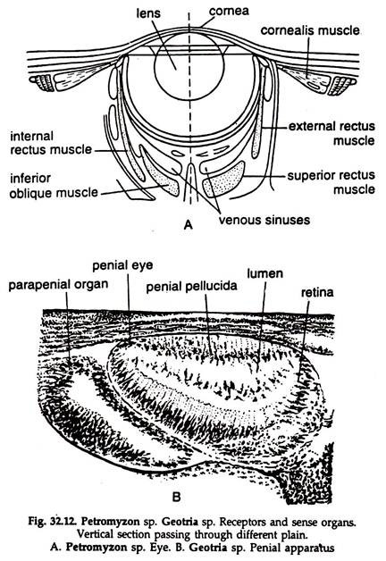
Eyes (Fig. 32.12) are almost similar to vertebrate eyes. The eye is attached to the rim of the orbit; the eye ball nearly spherical (Fig. 32.12A), cornea somewhat flattened, two-layered, separated by a gelatinous matrix; pupil round and capable of a little change in diameter. Extrinsic muscles move the eye-ball. Cornualis muscle attached to the outer layer of cornea helps in flattening the cornea and also in accommodation of the eye.
Pineal apparatus:
The pineal apparatus consists of a larger, dorsally placed pineal eye and a ventral, smaller parapenial body (Fig. 32.12B). Both develop from evagination of the roof of the brain and are connected with epithalamus.
The pineal eye is joined with the right Habenular ganglion by a stalk containing nerve fibres and the Para pineal body to the left Habenular ganglion with a similar stalk. Pigmented retina with sensory cells and a flattened imperfect lens are present in the pineal eye.
The structures are extremely reduced in Para pineal body. In lamprey, the pineal eye is better developed than that in any other vertebrate. The pineal eye is photosensitive and possibly helps in change of colour by sending impulses to the pituitary gland via hypothalamus.
Ear:
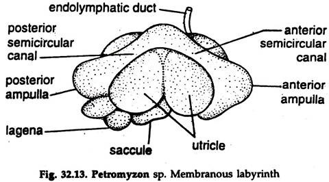
The auditory organs are represented by internal ears or membranous labyrinths (Fig. 32.13), nearly similar to those in other vertebrates. Vertical semicircular canals are two—anterior and posterior; bear ampulla at lower end containing crista ampullaris. An endolymphatic duct is present. A slight constriction indicates separation of utricle and saccule.
Lateral line sense organs:
The lateral line sense organs are simple and exposed to the surrounding water. They occur in the head region, extend in a single line along the body on each side and consists of small patches of sensory cells innervated by branches of fifth and tenth nerves.
Integumentary photoreceptors:
Occur in the tail of the ammocoete larva. They are supposed to be present in the skin of other parts of the body.
Gustatory receptors:
Organs of taste occur in the wall of the pharynx between gill pouches.
9. Reproductive System of Lamprey:
The sexes are separate in the adult. Both spermatocytes and oocytes may be found in a single gonad in young lamprey. Sexual differentiation occurs at a late stage.
The single gonad, testis, made of sperm follicles or ovary (Fig. 32.9) extends the length of the pleuroperitoneal cavity, remaining suspended from the mid-dorsal body wall by a membrane, the mesorchium or mesovarium in male and female, respectively.
Mature sperms or ova escape to the coelom with the rupture of the gonad and find their way through the genital pores to the urinogenital sinus and, thence, to the exterior.
Fertilization and development:
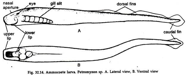
In the breeding season the adults migrate from sea to river. During spawning the male wraps the posterior part of the female body and sheds sperms over the eggs released by the female. Fertilization is external. The egg is telolecithal with considerable amount of yolk in the vegetal pole. At about twenty-one days, young Ammocoete larva hatches out.
Ammocoete larva:
It is dissimilar to adult:
1. Body small, about 10 cm long, trims- parent, with a median, continuous (Fig. 32.14) fin.
2. Instead of a suctorial buccal funnel of the adult a hood-shaped upper lip overhangs the mouth.
3. Teeth absent.
4. Velum, a pair of cup-shaped structures are attached to the anterior wall of the pharynx.
5. Entrance of the alimentary canal is guarded by a set of branched buccal tentacles.
6. Rudimentary paired eyes are hidden below skin and muscles.
7. Unpaired pineal eye well-developed.
8. Photoreceptors are abundant in the tail region.
9. The endostyle opens into the pharynx by a slit-like aperture and connected with hyper pharyngeal groove by a pair of peripharyngeal grooves.
10. The endostyle secretes a profuse quantity of mucus to which food particles are entangled. Cilia in the pharynx drive strands of food-laden mucus to the oesophagus and from there to intestine by oesophageal cilia.
11. The larva remains buried in mud in a U-shaped burrow, coming out only in night in search of food. It swims head downward and burrows again rapidly.
10. Metamorphosis in Lamprey:
The larval period is long and may extend for 3 to 4 years. Metamorphosis usually occurs in winter and the larva assumes the adult form.
1. Endostyle transforms into endocrine thyroid gland below the pharynx.
2. Suctorial buccal funnel with tongue, teeth and musculature surrounds the mouth.
3. The eyes complete their development and move to the surface.
4. The continuous dorsal fin changes to form two dorsal and a single caudal fin.
5. The oesophagus and respiratory tube turn to independent canals.
6. The velum reduced to guard the opening of the respiratory tube only.
7. The gills develop in sacs, the gill pouches opening in respiratory tube.
8. Nasal sac becomes folded internally and retains connection with enlarged olfactory sac.
9. The nasopharyngeal sac becomes enlarged posteriorly.
10. The larval cranium and branchial skeleton transform into the adult condition.
11. The skeleton of the upper lip and tongue develop.
12. Pronephros degenerates and mesonephros form the adult kidneys.
13. The spinal cord turns flattened.
14. The skin colour changes from muddy- brown to metallic on the dorsal surface.

