Here is a term paper on ‘Elphidium’. Find paragraphs, long and short term papers on ‘Elphidium’ especially written for college and medical students.
Term Paper # 1. Habits and Habitat of Elphidium:
Elphidium crispum represents a shelled-protozoan or framiniferan having many chambered shell and thread-like axopods. It exhibits the phenomenon of dimorphism, i.e., Elphidium exists in two distinct forms—large megalospheric and small microspheric forms. Elphidium is a marine and free-living protozoan. It is found on the bottom of sea at about the depth of 300 fathoms (one fathom = 1.8 metres). It creeps among the sea weeds.
Term Paper # 2. Morphology of Elphidium:
i. Shell:
Body of Elphidium is covered with a hard and translucent shell made up of calcium carbonate and small amounts of silica and magnesium sulphate. The shell is biconvex, polythalamus or multilocular (many chambered) and perforated. The surface of the shell is chiseled.
ADVERTISEMENTS:
The chambers of the shell are V-shaped, lay down serially and arranged in a flat spiral in which each whorl of chambers overlaps the previous whorl i.e., equitant. The overlapping portions are known as alar processes. Due to the overlapping of the chambers only the last chamber is visible from outside. The hinder margin of each chamber has a row of numerous minute backwardly directed, hollow, blind protoplasmic pockets called retral processes.
The adjacent chambers remain separated from each other by perforated septa. The chambers are interconnected or communicate with each other as well as with the exterior through minute pores present in the septa. The outer whorl opens to the outside by a row of large pores.
The chambers of the shell originate from the initial chamber known as proloculum which may be small or large in size. The small production is known as microsphere and the shell having small proloculum shall be called microspheric whereas the large proloculum is known as megalosphere and the shell having large proloculum is called megalospheric. Thus the animal is dimorphic.
ii. Cytoplasm:
ADVERTISEMENTS:
The shell’s chambers are filled with inner cytoplasm or medulla. A thin layer of outer cytoplasm or cortex covers the shell from outside. The inner cytoplasm contains nucleus or nuclei, food particles, minute vacuoles, Golgi apparatus, mitochondria, endoplasmic, reticulum, ribosomes and brown granules or xanthosomes containing waste matter. Contractile vacuoles are absent.
iii. Nucleus:
The medulla contains a single nucleus in megalospheric individuals and many nuclei in microspheric forms.
iv. Reticulopodia:
ADVERTISEMENTS:
The pseudopodia of Elphidium are in the form of numerous fine and often very long slender thread-like structures, which are often branched and anastomosing. This type of pseudopodia is characteristically called reticulopodia, rhizopodia or myxopodia. Each rhizopodium is made of an inner fibrillar axis and the outer fluid-like cortex.
The streaming circulation of cytoplasm has been observed in the rhizopodia. These are, in fact, temporary extensions of the outer cytoplasm and can be withdrawn within the shell. However, these are locomotory in function and often form feeding nets for catching diatoms on which animal feeds.
Term Paper # 3. Physiology of Elphidium:
i. Locomotion:
Elphidium creeps slowly with the help of its reticulopods on sea weeds at the bottom of the ocean. Locomotion of animal is performed by contraction and expansion (extension) of the reticulopods.
ii. Nutrition:
Nutrition is holozoic. The food consists mostly of diatoms and algae; it also captures other Protozoa and micro-crustaceans. The net-like rhizopodia are said to secrete an external mucus layer to entangle the food particles. The mucus layer also contains proteolytic secretions which help in paralysing the prey and the process of digestion soon starts.
The entangled food in mucus is enclosed in a food vacuole and then the rhizopodia are withdrawn within the shell. The food is digested almost exclusively outside the shell and the digested products pass into the inner cytoplasm.
Term Paper # 4. Reproduction and Life-History of Elphidium Crispum:
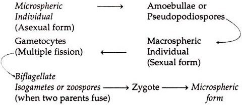
Elphidium multiplies by process of multiple fission and there is alternation of generations. The made of reproduction in different species of Elphidium is not exactly similar. The following is the description of life-history of Elphidium crispum.
Formation of Macrospheric Individuals:
ADVERTISEMENTS:
Microspheric form contains several nuclei. The smallest form as described by Schaudin had nine chambers and 28 nuclei. At first, nuclei are homogeneous structures, but they make their appearance as soon as the animal grows in size.
Nuclei are irregularly scattered, although they are absent in terminal chambers. Nuclei multiply by simple division. Nuclei in larger chambers are larger than those in the smaller chambers near the centre. Nuclei give off irregular strands of darkly stranding substances, which are termed by some authors as chromidia. In some cases, no definite nucleus is visible, only stained substance is present in the form of irregular strands.
The first indication of reproduction starts with the great increase in the number of pseudopodia. They become so abundant that they form a sort of a milk-while halo about the brown shell. This halo consists of hyaline protoplasm. Within a short time, coarse brown granules termed are chromidia pass out.
Whole of the protoplasm comes out of the test and is amassed within the area covered by the halo. Protoplasm by streaming movements separates into spherical masses of a uniform size; the centre of each is occupied by a nucleus with an area of clear cytoplasm surrounding it.
After a short time, each sphere or amoebula or pseudopodiospora thus formed secretes a shell. Each pseudopodiospore forms a number of chromidial granules, which fuse to form a nucleus. Soon, a second chamber is formed. Amoebula or pseudopodiospore grows, feeds and gives rise to a series of new chambers. Thus, a macrospheric form of Elphidium is formed.
Whole protoplasm in specimens fairly advanced in growth, Schulze noticed them in or near the chamber, which is almost in the middle of the series. Nucleus is at first a homogeneous structure, but soon it grows and round nucleoli are formed. Lankester describes that small fragments of irregular bodies are separated off from the nucleus.
Towards the end of the vegetative phase of the life-history of megalospheric form the nucleus disappears and minute nuclei are formed scattered uniformly or in groups throughout the protoplasm. When nuclei are uniformly distributed the protoplasm breakup into small rounded masses, the centre of which is occupied by a nucleus.
These nuclei divide by Karyokinesis or mitotic division. This is followed by a second division of protoplasm so as to from rounded bodies 3-4µ, termed as biflagellate isogametes, which are set free and escape outside the shell.
These gametes or zoospores from the same parent will not unite with one another; those from different parents will conjugate readily. Schaudin describes that the nuclei of the two gametes fuse and flagella drop off and the zygote thus formed undergoes a considerable increase in size, so that in a few hours its diameter is more than doubled.
The zygote secretes a gelatinous envelope and the microspheric individual is formed. Nucleus of the zygote divides by successive mitotic division and thus acquires multinucleate condition.
There is present alternation of generations in the life-history of Elphidium. Microspheric form gives rise to several amoebullae or pseudopodiospores, which form macrospheric forms. These in turn, give rise to biflagellate isogametes by multiple fission, which fuse and form zygotes. These zygotes form microspheric individuals and thus two generations alternate each other.
Life-History Formula of Elphidium:
