The nervous system of toad includes:
(1) The central nervous system,
(2) The peripheral nervous system, and
(3) The autonomic nervous system.
ADVERTISEMENTS:
The nervous system controls and co-ordinates the various activities of the body.
(1) Central Nervous System:
The central nervous system includes the brain and the spinal cord. It is essentially a hollow tube whose anterior portion becomes the brain and the posterior part narrows down to form the spinal cord. The cavity of the brain and the spinal cord is filled up with the cerebrospinal fluid.
The brain and the spinal cord are made up of nerve cells (neurons) and nerve fibres. A collection of the extended processes of the neurons enclosed by a connective tissue sheath constitute the nerves.
ADVERTISEMENTS:
Structure of a neuron:
A neurons has a very large cell-body with a number of processes. The nucleus of the cell- body is very conspicuous and the cytoplasm contains numerous deeply stainable granules called Nissl granules. The processes are usually branched, short and are called dendrites. One of the cell-processes is long and un-branched. It is known as axon which forms the axis cylinder of the nerve fibre.
The solid part of the central nervous system is composed of grey matter and white matter. The aggregation of nerve cells with their nuclei gives a greyish appearance and is recognised as the grey matter, while the collection of nerve fibres giving a white colour is known as the white matter.
The relative arrangement of grey matter and white matter differs in different regions. In brain, the grey matter is situated on the outer side and the white matter lies towards the lumen.
ADVERTISEMENTS:
But the arrangement is just reverse in the spinal cord, i.e., the grey matter is situated on the inner side and the white matter on the outer side. The whole of the central nervous system is enclosed by two protective coverings or meninges. The outer one is the Dura mater which is thicker and fibrous in nature. The inner one is thin, highly vascular and called pia mater.
Development of Central Nervous System:
The nervous system develops from the embryonic ectoderm. Along the mid-dorsal line of the whole length of the body above the notochord, the ectoderm forms a thickened neural plate. The sides of the neural plate rise up as lateral folds to form a neural groove. As development goes on, the edges of the groove fuse along the mid-dorsal line and is converted into a neural tube.
The anterior part is transformed into the brain and the posterior part forms the spinal cord. The brain was primarily a tube-like structure, which, due to unequal growth, torsions and bending, transforms into a most complicated adult structure.
Brain:
The brain is located inside the cranium. It is primarily differentiated into three parts by the development of two primary constrictions: the forebrain (prosencephalon), the midbrain (mesencephalon) and the hind- brain (rhombencephalon). The prosencephalon constitutes the anteriormost part of the brain and becomes further subdivided into an anterior telencephalon and a posterior diencephalon (thalamencephalon).
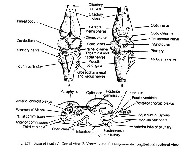
The telencephalon gives off a pair of small olfactory lobes. These lobes are responsible for the sense of smell. The rest of the telencephalon consists of two elongated ovoid lobes (Fig. 1.74A) called cerebrum (cerebral hemispheres). The roof of the cerebrum is very thin and the floor is very thick and called corpus striatum.
Two corpora striata are connected together by an anterior commissure. Above this commissure, there is an ill-developed pallial commissure. The cerebrum is the seat of consciousness, intelligence and it regulates voluntary motion. The surface of the cerebral hemispheres is smooth.
The diencephalon is a very depressed part behind the cerebrum. It gives off dorsally a small projection — the epiphysis. Attached to the epiphysis, there is a small pineal body of unknown function. In front of the epiphysis there is a vascular membrane projecting into the lateral ventricle through the foramen of Monro.
ADVERTISEMENTS:
This projection is known as anterior choroid plexus. The ventral side of the diencephalon bears an X-shaped optic chiasma. The optic chiasma is formed by the optic nerves (Fig. 1.74B). Just a little behind the optic chiasma hangs a projection, the hypophysis (or infundibulum). The pituitary gland is attached at the tip of the infundibulum.
The lateral sides of the diencephalon become thick to form the optic thalami. The midbrain remains undivided and grows out dorsally as a pair of hollow ovoid bodies known as corpora bigemina (or optic lobes). Two longitudinal bands are present on the ventral sides which connect the forebrain with the hindbrain. These are designated as crura cerebri.
The rhombencephalon is the posteriormost sector of the brain and becomes divided into metencephalon and myelencephalon. The metencephalon is represented by a thin transverse band-like cerebellum. The floor and the sides of the myelencephalon become very thick to form the medulla oblongata.
The cerebellum coordinates the movement of the body. The medulla oblongata controls and regulates some of the vital processes, viz., the regulation of heart beat, metabolism and respiration. The roof of the myelencephalon is formed by a thin vascular membrane called posterior choroid plexus.
Cavities in brain:
The brain is not a solid structure but contains well-formed internal cavities called ventricles. The cavities in the cerebral hemispheres constitute the lateral ventricles (the first and the second ventricles). The cavity in the diencephalon constitutes the third ventricle and that of medulla oblongata is the fourth ventricle.
The lateral ventricles remain in communication with the third ventricle by a small opening called foramen of Monro. The third ventricle communicates with the fourth ventricle by a narrow passage (Fig. 1.74C) called aqueduct of Sylvius (iter). The fourth ventricle leads into the cavity of the spinal cord. The cavity in the optic lobes is known as optocoel and that in the olfactory lobes as rhinocoel.
Spinal cord:
The spinal cord is a hollow tube. It extends posteriorly from the medulla oblongata and remains encased within the neural canal of the vertebral column. It extends even within the urostyle as a slender filament, the filum terminale.
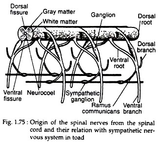
The spinal cord has a mid-dorsal groove called dorsal fissure and a similar ventral fissure is present on the mid-ventral line (Fig. 1.75). The cavity of the spinal cord is known as neurocoel which is also continuous with the ventricles of the brain.
(2) Peripheral Nervous System:
The peripheral nervous system includes the cranial and the spinal verves arising from the cerebrospinal axis. A nerve is a bundle of nerve fibres, but in some cases, an aggregation of nerve cells causes swelling on the nerve fibre.
This swelling is called ganglion. The nerve fibres are of two types — afferent or sensory fibres convey the information from the receptor organs to the central nervous system, and efferent or motor fibres carry impulses from the central nervous system to the effector organs.
When the nerves are exclusively made up of sensory nerve fibres, these are called sensory nerves and those composed only of motor nerve fibres are known as motor nerves. Mixed nerves are also present which are composed of sensory as well as motor nerve fibres.
Cranial Nerves:
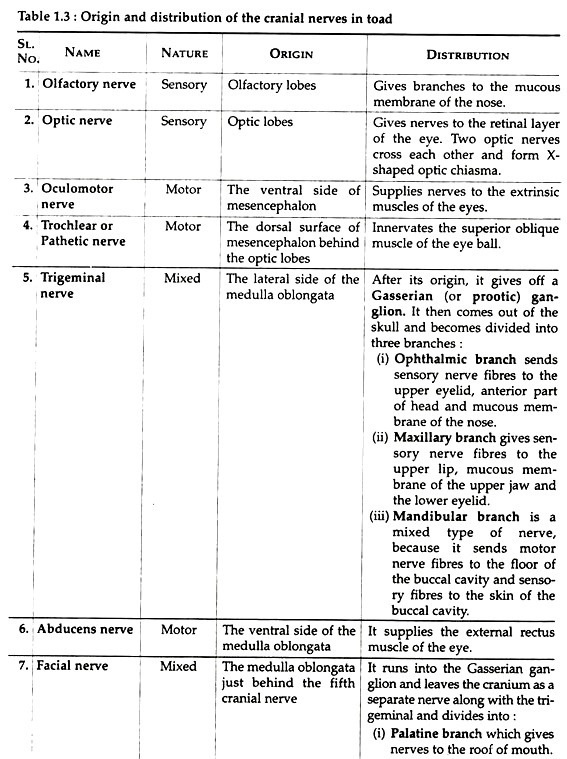
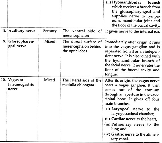
Ten pairs of cranial nerves originate from the brain. These paired nerves are designated as cranial nerves. Table 1.3 will give an idea about the origin and distribution of the cranial nerves in toad.
Spinal Nerves:
There are ten pairs of spinal nerves in toad. All the spinal nerves in toad are of mixed in nature. Each spinal nerve has a dorsal sensory root and a ventral motor root. The dorsal root possesses a ganglion near its origin. The dorsal and ventral roots unite and form a common trunk which comes out through intervertebral foramen and gives off three branches (Fig 1.75):
(1) A large ventral branch which innervates the skin and muscles on the ventral surface of the body.
(2) A small dorsal branch which supplies the skin and muscles on the dorsal side of the body.
(3) A very small ramus communicans which joins with the nearest sympathetic ganglion.
The first spinal nerve is known as hypoglossal. It supplies the muscles of the tongue. The second and third spinal nerves form the brachial plexus. The fourth, fifth and sixth spinal nerves supply the integument and the muscles of the trunk region. The rest of the spinal nerves form the sciatic plexus which gives orgin to the large sciatic nerve and supply the hind limb. The last spinal nerve is insignificant.
(3) Autonomic Nervous System:
The autonomic nervous system consists of two sympathetic trunks one on either side of the dorsal aorta. The sympathetic trunks start near the point of formation of iliac arteries and each trunk contains ten small ganglia connecting with the corresponding spinal nerve by ramus communicans.
Anteriorly, the sympathetic trunk divides to encircle the subclavian artery. It then enters into the cranium, connects by a twig with the vagus ganglion and terminates after connecting with the Gasserian ganglion. The sympathetic trunks give out branches to innervate the cardiac muscles, blood vessels and alimentary canal.
These branches may sometimes unite to form ganglionated plexuses, such as the cardiac plexus and solar plexus. The autonomic nervous system regulates the involuntary activities of the body, like heart beats peristalsis of the alimentary canal etc. It is called autonomic nervous system in the sense that its activities are somewhat independent of the central nervous system.
Sense Organs:
The sense organs are the receptors for external stimuli. These are the avenues through which the central nervous system is kept informed of the outside world. Each receptor organ can respond to a particular type of stimulus and produces its own specific sensation. Receptors for cold, heat, pain and touch are present beneath the epidermis of the skin. These are microscopic in structure and are usually regarded as cutaneous receptors.
Receptors for smell:
These receptors are scattered in the nasal passage. The mucous membrane, lining the nasal passage contains peculiar olfactory cells and slender supporting cells. The olfactory cells are connected with nerve fibres from the olfactory nerve and are the actual receptors for smell.
Receptors for taste:
The taste buds present in the tongue and mouth cavity are the receptors for taste. Each taste bud is made up of two types of cells — the taste cells and the supporting cells. The taste buds are located in the tongue in association with minute elevations called papillae.
Receptors for vision:
The eyes are the two very compact photosensitive organs. These organs are lodged in the orbits. Each eye has a spherical body and is usually called eye ball.
The eye ball can be rotated inside the orbit within its limit by six extrinsic muscles, viz.:
(1) Superior rectus,
(2) Inferior rectus,
(3) External rectus,
(4) Internal rectus,
(5) superior oblique, and
(6) Inferior oblique.
The eye can be protruded by the levator bulbi and can be withdrawn by the retractor bulbi to a certain limit.
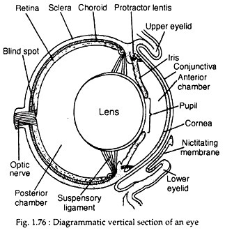
The eyeball is composed of three layers arranged in a regular sequence (Fig. 1.76).
(a) Sclerotic layer or Sclera:
This is the outermost layer of the eye ball. It is very tough and thick. It consists of two parts – an anterior transparent circular portion called cornea and an opaque posterior portion called white of the eyeball. The cornea permits the entry of light rays inside the eye. The outer surface of the cornea is covered by a thin transparent membrane called conjunctiva. The sclera is composed of cartilage and fibrous tissue and gives protection to the delicate parts of the eye.
(b) Uvea or middle layer:
This layer is divided into three parts:
(i) Choroid,
(ii) Iris, and
(iii) Suspensory ligament.
The choroid part is a highly vascular pigmented layer. At the anterior end and just behind the cornea, the choroid is modified to form a circular pigmented disc called iris. The iris contains an aperture at the centre which is known as pupil.
The iris acts as a diaphragm and contains circular and radial muscle fibres which help the pupil to contract or dilate. Excepting the pupil, the eyeball is totally light-proof. The iris regulates the entry of light into the eyeball. Just behind the iris, there are suspensory ligaments which keep the lens in position.
(c) Retinal layer:
The retinal layer forms the innermost and the light sensitive screen of the eye. It contains two types of photosensitive cells called rods and cones. The cones are primarily concerned with the colour vision in bright light and the rods are chiefly useful in colourless vision at low light intensities. The photosensitive cells are connected with the nerve fibres of the optic nerve.
A crystalline lens is situated just behind the pupil and is kept in position by suspensory ligament. The lens divides the cavity of the eye into two chambers. The anterior chamber between the lens and the cornea is filled up with a watery fluid, the aqueous humor and the posterior one behind the lens is filled up with a transparent jelly-like substance, the vitreous humor.
Just at the point of entry of the optic nerve into the retinal layer there is a depression known as blind spot where no image is formed. Two muscles, one dorsal and the other ventral, are connected with the suspensory ligament of the lens and with the cornea. These muscles are known as protractor lentis. Contraction of the protractor lentis draws the lens closer to the cornea and when relaxed the lens is pushed away from the cornea.
Receptors for hearing and balancing:
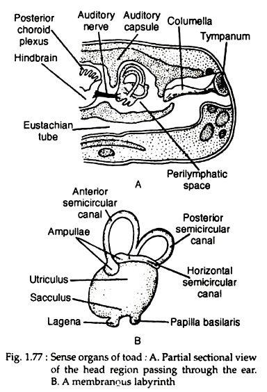
The ears sub serve dual functions, hearing and balancing. The ear of toad consists of three parts: the external ear, the middle ear and the internal ear (Fig. 1.77A). The external ear is represented by a tightly stretched membrane called tympanum. The middle ear is a tube like cavity. The cavity of the middle ear is in communication with the buccal cavity by eustachian tube.
Presence of the eustachian tube equalises the atmospheric pressure on the two sides of the tympanum. A bony rod, the columella connects the tympanum with the membranous partition separating the middle and the internal ear. The internal ear is represented by the membranous partition separating the middle and the internal ear. The internal ear is represented by the membranous labyrinth which is enclosed by the auditory capsule (Fig. 1.77A).
The membranous labyrinth floats in a fluid known as perilymph and the cavity of the labyrinth is filled with another fluid known as endolymph. The auditory capsule is sealed from all sides by a membranous partition. Membranous labyrinth is made up of two chambers. The upper chamber is called utriculus and the lower one is the sacculus.
The utriculus gives out narrow tubular semicircular canals (Fig. 1.77B). There are three semicircular canals, one is horizontal in position and the other two are vertically disposed. All the three semicircular canals are arranged at right angles with one another. Both the ends of the semicircular canals open into the utriculus.
Each canal bears an ampulla at one end only. The sacculus produces a short projection called lagena. Patches of sensory receptors are present in the inner wall of the membranous labyrinth. Each patch is composed of sensory cells and supporting cells. Receptor cells are connected with the nerve fibres from the auditory nerve.
Mechanism of hearing:
Sound waves directly impinge upon the tympanum and the vibrations are conveyed by the columella to the perilymph. From the perilymph, the vibrations are carried to the endolymph and thus stimulate the sensory cells of the sacculus and lagena. The impulses are transmitted to the brain through the auditory nerves and are perceived as sound in the brain.
Mechanism of balancing:
The semicircular canals maintain the equilibrium of the body. Calcareous particles (otoliths) present inside the semicircular canals strike upon the bristles of receptor as the animals lose the balance. Besides, the semicircular canals are arranged in such a fashion that these can easily detect changes of the centre of gravity during movement.
