The embryology of chick or the common fowl (Gallus gallus) is preferable to study to that of pigeon (Columba livia) because of several advantages.
The embryology of chick has been worked out more extensively only because of the following reasons:
1. Eggs of fowl are large or of convenient size, easily available throughout the year and can be incubated artificially.
2. The various developmental stages can be easily available in laboratory conditions for experimental purposes.
ADVERTISEMENTS:
3. Moreover, the process of development has been most thoroughly worked out in fowl.
4. The embryology of chick bears many resemblances with those of reptiles and mammals. A comparative study of embryology of different birds exhibits that it is essentially similar in all the birds with only minor unimportant differences. Despite these reasons, the embryology of chick is important due to phylogenetic significance. In chick, development is direct without a larval stage.
Fertilisation:
The ova are released from the ovary in the form of primary oocytes. The second maturation division occurs after ovulation in the oviduct. The released primary oocyte in the body coelom is grasped and swallowed by the ostium of the oviduct.
The secondary oocyte or ovum after second maturation division is surrounded by several (5 or 6) sperms which enter in it (polyspermy), but only one sperm succeeds in the fertilisation process. The nucleus of one sperm fuses with the female nucleus (amphimixis) and the nuclei of other sperms degenerate.
ADVERTISEMENTS:
As the fertilised egg passes downward inside the oviduct, it rotates, undergoes cleavage and various accessory egg membranes are laid down over the developing egg. Thus, fertilisation is internal. The various egg membranes secreted around egg are albumen, shell membranes and shell.
Structure of Egg of Hen:
The egg is about 3.0 cm in diameter and is polylecithal. It is entirely filled up by the yolk and over it, i.e., in animal pole lies a small cytoplasmic disc with a nucleus. This disc is called blastodisc. The yolk has a central mass of white yolk around which are alternate concentric layers of yellow and white yolk. The central flask-shaped white yolk called latebra runs from the centre to the lower side of the blastodisc and there it spreads to form the nucleus of Pander.
The yolk and blastodisc are bounded by a plasma membrane and outer vitelline membrane, which is a converted form of zona radiata. The vitelline membrane is of double origin. The inner layer of this membrane is produced in the ovary between the oocyte and follicle cells and it is composed of very tough fibres. The outer layer of it is formed in the upper part of fallopian tube.
The yolk contains 48.7% water, 32.6% phospholipids and fats, 16% proteins, 1% carbohydrates and 1.1% other chemical molecules. Its proteins are in the form of phosvitin and lipovitelline or livetin. The fat is predominantly neutral fat (50%), rest is phosphatids, cerebrosides and cholesterol.
The fertilised egg or zygote is covered by a layer of dense viscous albumen which forms a thin chalaziferous layer around the vitelline membrane. This dense albumen forms two twisted cords or chalazae, one at each end of the zygote. They are formed by rotation of the egg during its movement through the oviduct.
Around the chalaziferous layer is a thick layer of watery albumen. All albumen is secreted by the upper glandular walls or magnum of the oviduct. The functions of albumen are to provide nutrition to the embryo, serves as a water store and also acts as protective envelope for protecting the embryo from mechanical and chemical injuries.
The albumen and yolk also contains a variety of enzymes, vitamins, pigments and phosphorus. The isthmus part of the oviduct secretes two shell membranes made of tough keratin fibres matted together. The two shell membranes are closely applied except at the blunt end of the egg where they are separated by an air space formed after the egg is laid. The nidamental glands of the oviduct secrete a porous, calcareous shell which soon hardens.
It is pierced by a large number (about 7,000) of fine pores filled with a protein related to collagen. The diameter of the pores varies from 0.04 to 0.05 mm. These pores allow exchange of gases (oxygen and carbon dioxide) during respiration of the developing embryo. The egg is laid 24 hours after fertilisation, and its further development takes place, when the egg is incubated by the female. Incubation must continue steadily for 21 days at a temperature of 103°F.
Cleavage and Blastulation:
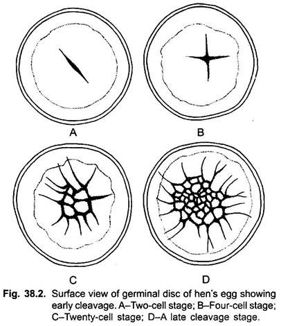
Cleavage is meroblastic or discoidal and is confined only to be germinal disc or blastodisc. It does not segment the yolk of the polylecithal egg and it is later eventually surrounded by the growing tissues of the embryo. First two cleavages are at right angles to each other in the centre of blastodisc. These cleavage furrows do not cut the germinal disc completely through in the vertical plane.
The third set of cleavage furrows is vertical, cutting across the second set of vertical furrows. The fourth cleavage furrow is also vertical and circular cutting across all the cleavage furrows, forming eight central blastomeres which are surrounded by eight marginal blastomeres. Thus, these cleavage furrows separate the daughter central blastomeres from each other, but not from the yolk. The central blastomeres are continuous with the underlying yolk at their lower ends.
The marginal blastomeres are continuous with the uncleaved cytoplasm at their outer edges. Further cleavages are irregular. The central cells divide more rapidly. The marginal cells also divide by the appearance of new horizontal and radial furrows. The newly formed inner cells of marginal blastomeres are added to the central cells, resulting in the increase of volume of this area. The radial furrows extend peripherally and these peripheral cells are still continuous with the uncleaved peripheral cytoplasm.
In later stage of cleavage, the blastomeres of the central area become separated from the underlying yolk due to the appearance of a horizontal cleavage in these cells. This cleavage extends peripherally cutting the inner ends of the blastomeres. Thus, a space also appears in the beginning beneath the central cells which also extends peripherally as the horizontal cleavage extends outward.
ADVERTISEMENTS:
The cavity beneath the central cells, i.e., in between central cells and yolk is called the subgerminal cavity, which is filled with a fluid diffused from the albumen through vitelline membrane. Thus, due to further cleavage the blastodisc becomes cellular, called the blastoderm- a round disc, 5 to 6 cells deep in the centre but only 1 to 2 cells deep at the periphery.
The appearance of subgerminal cavity separates the blastoderm from the underlying yolk, but the marginal cells remain overlapping the yolk. The embryo is now called the blastula stage. At blastula stage the embryo reaches in the uterus. It may be compared to the blastula of Amphioxus and frog, its sub-geminal cavity is equivalent to the blastocoel, the blastoderm is the animal pole, and the yolk is the vegetal pole.
During later part of cleavage, about 12 to 14 hours after the egg reaches the uterus or 6 to 8 hours before the egg is laid, some cells on the inner or under side of the blastoderm become detached or delaminated from the blastoderm and fall on the floor of subgerminal cavity due to presence of relatively more yolk. The delamination of these yolky cells from the blastoderm starts at the posterior edge and spreads forward until whole blastoderm becomes free from yolky cells.
As a result, the epithelial layer in the central region of blastoderm becomes thinner (few layers of cells) and transparent. Thus, this region is called the area pellucida because it seems to be transparent when viewed from the upper side. The peripheral part of blastoderm, the yolky cells is not delaminated (shed), so this part of the blastoderm seems to be opaque, because beneath these cells blastocoel is not present.
This region of blastoderm is, thus, called area opaca. These delaminated cells at the posterior edge of area pellucida gradually link up with each other, forming a, continuous layer of flattened cells, which extends anteriorly. This layer is called the hypoblast and the upper layer is the epiblast containing ectoderm and mesoderm cells. Hypoblast is exclusively composed of endoderm cells.
The egg is laid by the female about the time the blastula is formed or even a little later.
Presumptive Fate Maps of Blastula:
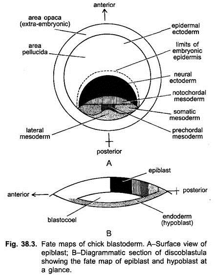
Fate maps of the blastula of chick have been prepared by using the vital stains such as carmine or carbon (charcoal) particles or radioactive thymidine. It shows that blastomeres of area opaca do not form any part of the embryo proper, they form only extra-embryonic membranes.
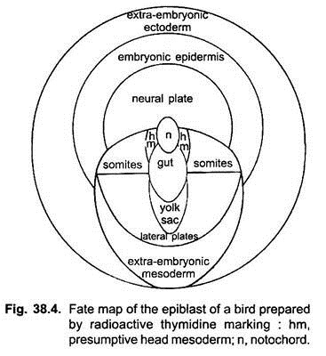
The epiblast and hypoblast of area pellucida have different fates in the course of embryonic development. The fate maps prepared by the use of tritiated thymidine have shown the following structures in epiblast- In the centre of area pellucida lies a small area destined to produce the notochord. Posterior to it, in the median plane is found an elongated oval area of presumptive endoderm which will form the gut.
Further toward the posterior edge of area pellucida lies the extra-embryonic endoderm which will form the lining of yolk sac. To the right and left of the presumptive notochord and endoderm, and posterior to extra-embryonic endoderm lie the various subdivisions of presumptive mesoderm, i.e., prechordal plate or head mesoderm, mesodermal somites, lateral plate mesoderm and extra-embryonic mesoderm.
The anterior half of epiblast is the presumptive ectoderm containing central presumptive neural plate area, anterior to it is the presumptive embryonic epidermis and outer to it in the form of complete ring is the extra-embryonic ectoderm.
Gastrulation:
Gastrulation includes the following two types of morphogenetic movements:
a. Emboly:
It involves only the epiblast which contains cells of ectoderm,; mesoderm and notochordal cells. It includes convergence, invagination and involution. Formation of primitive streak and head process is due to emboly.
b. Epiboly:
It includes the overgrowth of ectoderm or epiblast and also of hypoblast.
Formation of Primitive Streak:
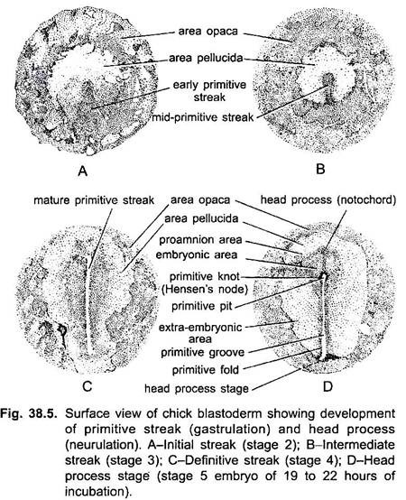
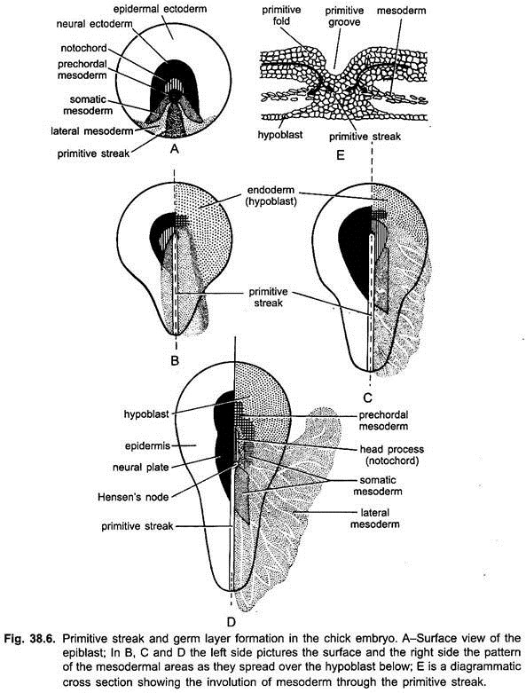
Various prospective mesodermal and endodermal cells forming notochord of the epiblast converge toward the posterior edge of the area pellucida and form a conical thickening in the midline, called the initial primitive streak. It appears after 6 to 7 hours of incubation.
The primitive streak grows anteriorly because of proliferation of its own cells as well as of the addition of cells that migrate to it from anterior and lateral parts of area pellucida. The elongated axis of the primitive streak marks the antero-posterior axis of the future embryo. It, thus, eventually extends to, about three fifths of the entire length of area pellucida.
This is the fully developed definitive primitive streak and it is usually completed after 18 to 19 hours of incubation. The area pellucida also becomes pear-shaped. Along the middle of the primitive streak, when it is fully developed runs a narrow furrow, the primitive groove. At the anterior end of the primitive streak there is a thickening, the primitive knot or Hensen’s node. The centre of Hensen’s node is excavated to form a funnel-shaped depression.
The movements in the blastoderm leading to the final placement of cells in the hypoblast and to the formation of the primitive streak in the epiblast may be called pregastrular movements.
Invagination and Involution:
At the stage of short primitive streak, the cells of the blastoderm already begin to migrate (invaginate and involute) into the blastocoel cavity between epiblast and hypoblast. Immigrating cells are replaced by more epiblast cells converging toward the streak area.
The inward migrating cells also spread out sideways and forward from the anterior end of primitive streak. The notochordal cells immigrate through primitive pit. Endodermal cells invaginate through that part of the streak which lies just behind primitive pit.
The mesodermal cells of somites just follow the path of endodermal cells. Whereas the lateral plate mesoderm cells invaginate through the middle section of primitive streak, but only after the disappearance of endoderm from the area pellucida. The extra-exbryonic mesoderm (of the yolk sac) immigrates through the posterior part of primitive streak.
Meanwhile, some hypoblast cells expand into the area opaca to become extra-embryonic endoderm (the lining of yolk sac), while other hypoblast cells attach to mesodermal and notochordal cells are carried along by the latter’s migration.
Formation of Head Process:
Prospective notochoral cells converge on the node, sinks through it and then passes directly forward as a tongue of tissue known as head process or notochord process.
Disappearance of Primitive Streak:
With the gradual disappearance of endodermal, notochordal and mesodermal cells from the primitive streak, it begins to shrink from anterior towards posterior side and its remains are partly included in the tail bud and partly into the cloacal region of the embryo.
Development of Head Process:
The midline area of notochordal tissue develops into a rigid rod, anterior to the receding primitive streak. As the streak regresses posteriorly, the embryo develops anterior to it. The head process consists of a thick central mass of cells and more diffuse lateral wings. In the beginning it is also blended in the midline with the hypoblast.
The thicker central portion forms the definitive notochord, whereas the lateral wings form the paraxial (somitic) mesoderm. With its differentiation, the notochord becomes detached from the hypoblast below, except at the extreme end. Thus, the head process stage is completed at about 20 to 25 hours of incubation. Gastrulation is also completed at this stage.
Completion of Endoderm:
The first cells that migrate through the anterior part of streak form the endoderm. As the Hensen’s node recedes backward, and the notochordal process elongates, the presumptive endoderm of the middle and posterior part of the gut, located just behind the node, migrate inside as an endodermal strip beneath the notochord.
The original hypoblast at the floor of the blastocoel contribute a very less amount to the gut, the upper migrated endodermal cells form the major part of the gut. In chick no archenteron is formed during gastrulation.
Fully Formed Gastrula:
Gastrula is fully formed when primitive streak completely disappears. The fully formed gastrula consists of three germ layers-ectoderm, chorda-mesoderm and endoderm. The ectoderm and chorda-mesoderm remain in continuity along the axis of primitive streak. The endoderm is also united with the mesoderm and ectoderm at the anterior and posterior end of streak.
Formation of Neural Tube (Neurogenesis):
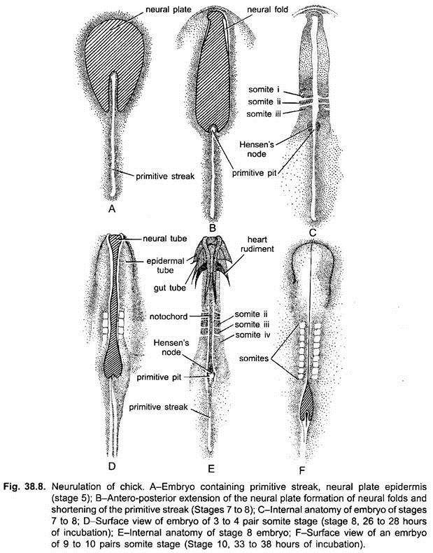
The ectoderm, anterior and lateral to the head process, becomes thickened to form the neural plate while the gastrulation process is going on. The neural plate appears in the brain region. As Hensen’s node recedes farther and farther, parts of the neural plate become differentiated, and the anterior parts of the neural plate proceed to close into a tube, the neural tube.
The formation of neural tube occurs due to sinking in of the neural plate due to which a neural groove is formed along its longitudinal axis. Its elevated margins are called neural folds, which rise up and grow toward the midline and fuse to form the neural tube.
Formation of Notochord and Mesoderm:
While the neural plate is folding into the neural tube, the chorda-mesoderm is also differentiating. Its most anterior part, the prechordal plate mesoderm gives rise to the mesenchyme of head, and behind it the notochordal cells become separated from the rest of the adjoining sheets of mesoderm and differentiated into notchord. On either side of notochord are three longitudinal strands or sheets of mesoderm.
1. Epimere, axial or somatic mesoderms are the thicker dorsal medial strands, which merge anteriorly into the mesenchyme of head.
2. Mesomeres or intermediate mesodermal strands are located lateral to the axial mesoderm. They are thin in an early stage.
3. Hypomeres or lateral plate mesoderm lies beneath the mesomere.
Formation of Somites:
The axial or somatic mesoderm begging’s to differentiate after at about 21 hours of incubation. It becomes a thick band on either side of notochord and nerve cord. A short distance anterior to the primitive knot a transverse cleft appears across each band, marking the division between first and second somites.
About one hour later a second cleft appears posterior to first cleft and, thus, second somite is formed. This process goes on approximately after each hour up to 20 hours. After that somites formation become somewhat slower.
The first three pairs of mesodermal somites are formed in the paraxial mesoderm in the presumptive hind brain region, and other mesodermal somites appear as the primitive knot regresses. Somites, when first formed, are masses of mesodermal cells with a small cavity in the centre. The cells are arranged radially around the central cavity.
With further development, they become extended in the dorso-ventral direction and flattened medio-laterally. The cavity (myocoel) of each somite also changes, from spherical to a narrow vertical slit. An inner wall and an outer wall become clearly distinguishable, corresponding to the parietal and visceral layers of the lateral plates. The inner wall of the somites becomes very much thicker and produces skeletogenous tissue and the voluntary striated muscles of the body.
The outer of the somites is thinner and forms the connective tissue layer of the skin and is, therefore, called the dermatome. Furthermore, the dorsal part of the inner wall of the somite which is the source of somatic muscles of the body is called myotome, whereas the lower edge of the inner wall is called sclerotome which is responsible for skeletogenous tissue. The sclerotome breaks up into a mass of mesenchymal cells.
The cells migrate into the spaces surrounding the notochord and spinal cord, envelop these organs and later differentiate into cartilage, thus, forming the bodies and neural arches of the vertebrae. Haemal arches and the ribs are also formed by these cells.
Formation of Coelom:
After the somites and the lateral plates have been formed, the mesoderm of lateral plate splits into two layers- the external or parietal layer and the internal or visceral layer. The cavity between the two is the coelom. Thus, the origin of coelom is schizocoelic.
Folding of Embryo:
The entire blastoderm does not form the embryo. Its central portion, called the area pellucida, only forms the embryo, and its outer portion, the area opaca forms the extra-embryonic membranes, such as yolk sac, amnion, chorion and allantois.
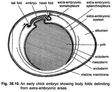
The body of the embryo becomes separated from the yolk sac. This is achieved by the formation of folds which appear all around the body of embryo. The folds involve all three germ layers and are directed downward and inward, undercutting the body of the embryo proper. They are known as body folds. The various folds do not appear simultaneously.
The first to appear is the head fold just in front of the head. It undercuts the head and anterior part of the trunk of the embryo, so that these parts project freely over the surface of yolk sac. The lateral and posterior parts of the body fold develop soon after.
The posterior fold undercuts the tail end and the posterior part of trunk which also project freely over the surface of yolk sac. These body folds gradually contract underneath the embryo and eventually the body of embryo is connected with the yolk sac and other extra-embryonic membranes by a narrow stalk, the umbilical cord.
Flexure and Torsion:
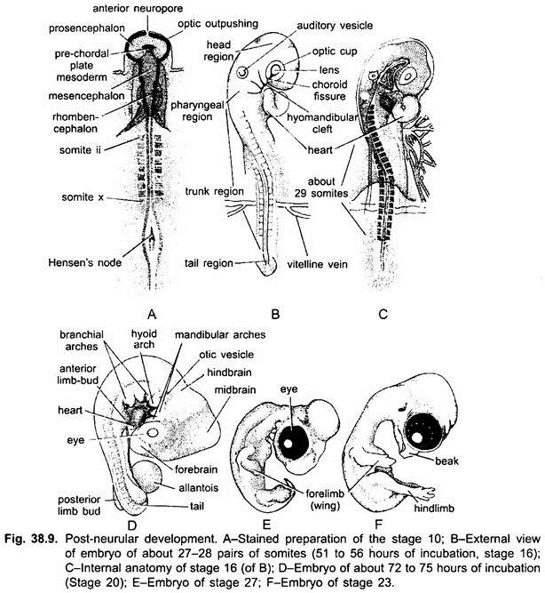
When the head is formed it gets bent down ventrally with respect to the main axis of the embryo, this bending is called cranial flexure. Then the head at first, and gradually the entire body turn sideways so that the embryo comes to lie on its left side over the yolk, this dextral twist is knows as torsion. The anterior end in its cervical region becomes still more bent over towards its ventral surface.
Various organs are formed on the third and fourth days of incubation, such as the nervous system, sense organs, four pairs of pharyngeal pouches out of which only the first three pairs meet the ectoderm to open as gill-clefts, five pairs of visceral arches, heart and, blood vessels, and paired segmental somites.
Extra-Embryonic (Foetal) Membranes of Chick:
The blastoderm besides forming the embryo, gives rise to certain other structures which do not take part in the formation of embryo proper, but are external to the developing embryo. These structure are collectively called foetal membranes, embryonic membranes or extra-embryonic membranes.
These membranes are essential for complete development of the embryo. These foetal membranes are concerned with nutrition, excretion, respiration and protection from desiccation and concussions. These foetal membranes are amnion, chorion or serosa, allantois and yolk sac.
These membranes are composite structures involving two germ layers. The amnion and chorion are composed of extra-embryonic ectoderm and somatic layer of mesoderm, both are collectively called somatopleure. Whereas the allantois and yolk sac are formed of extra-embryonic endoderm and splanchnic mesoderm layers, both are collectively called splanchnopleure.
Development of Extra-Embryonic Membranes:
During neurulation, the lateral plate mesoderm splits into an outer somatic layer lying beneath the ectoderm and an inner layer lying outer to the endoderm layer. It between these two layers of mesoderm lies the coelomic space.
The ectoderm and somatic layer of mesoderm are collectively called somatopleure, while the splanchnic layer of mesoderm and endoderm form the splanchnopleure. During later developmental stages, the somatopleure and splanchnopleure gradually spread outward beyond the developing embryo.
As the body of embryo grows, it becomes separated from the foetal membranes by the appearance of body folds-cephalic, caudal and lateral folds. These folds limit the body of embryo and separate the embryonic region from the extra-embryonic region.
1. Development of Amnion and Chorion:
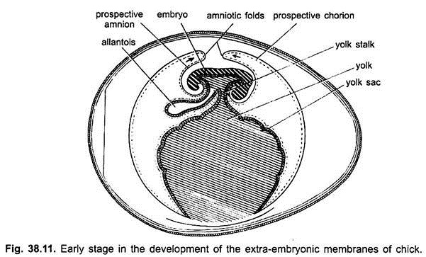
The origin of amnion and chorion is considered together since they develop simultaneously from the extra-embryonic somatopleure. About 30 hours of incubation, the extra-embryonic somatopleure of the blastoderm rises up in front of the embryo as a fold, the fold grows and forms an arch over the embryo.
It forms a double somatopleuric hood, called the caphalic amniotic fold. As this fold gradually extends backward, its caudally extending side limbs called lateral amniotic folds arch over the embryo from either lateral side. A similar fold or elevation over the tail appears, called the caudal amniotic fold. All these amniotic folds converge and fuse over the embryo, enclosing it within two sheets of somatopleure from all sides except the region of yolk stalk.
The region of union of the amniotic folds is called the seroamniotic connection and the scar-like point of fusion of the folds is called the seroamniotic raphe. The seroamniotic connection opens before the hatching of embryo. The fusion of amniotic folds produces two sac-like cavities, enclosed by two sheets of somatopleure.
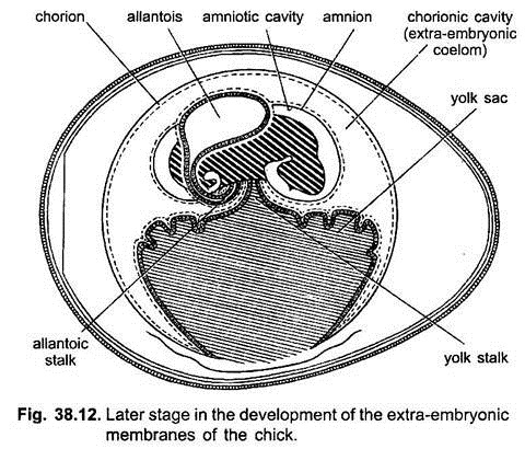
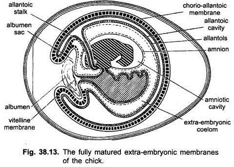
The inner somatopleuric sheet becomes the amnion, enclosing the embryo within amniotic cavity filled with amniotic fluid. While the outer sheet of somatopleure is the chorion and the cavity lying between the amnion and chorion is the chorionic cavity or extra-embryonic coelom. It is lined by mesoderm of somatopleure and splanchnopleure.
The somatopleure of amnion and chorion grows continuously peripherally and laterally and ultimately encloses the embryo and also the remaining two foetal membranes.
Functions of Amnion and Chorion:
The amnion, chorion and amniotic cavity serves the following functions for the embryo:
a. The amnion protects the embryo from desiccation and sudden temperature changes.
b. The amniotic fluid is an efficient shock absorber and protects the embryo from the mechanical -shocks.
c. The shell and shell membranes are protective and prevent desiccation of the embryo.
d. The mesoderm of amnion during later development forms muscle cells which contract rhythmically, rocking the embryo with in the amniotic fluid so as to prevent it from adhesion to the embryonic membranes.
2. Development of Allantois:
About the third day of incubation the floor of the endodermal hindgut begins to bulge as a bladder, called the allantois. The allantoic evagination is formed from the splanchnopleure, that is, inner layer of endoderm and an outer layer of splanchnic mesoderm. It grows rapidly and spreads into the extra-embryonic coelom, the space between the yolk sac, the amnion and the chorion.
The distal part of the allantois expands and remains connected with the hindgut of the embryo by means of a narrow allantoic stalk. When the body folds contract, separating the embryo from the extra-embryonic parts, the allantoic stalk is enclosed together with the stalk of yolk sac, forming an umbilical cord.
As the allantois vesicle enlarges and spreads outward, its distal part becomes flattened and expands between the amnion and yolk sac on one side and the chorion on the other side. Due to this the splanchnic mesoderm of the allantois fuses with the inner somatic mesoderm of the chorion to form an allanto-chorion.
The allanto-chorion grows around the albumen of the egg to enclose it in an albumen sac. It aids in absorption of water and albumen. The allantois becomes highly vascular due to the appearance of blood vessels in the splanchnopleure. It also receives a pair of allantoic or umbilical arteries, and its blood is returned into a pair of allantoic or umbilical veins.
The chorio-allantoic circulation continues until the young chick breaks the egg-shell and begins to breathe the surrounding air. Thus, the umbilical vessels close, the circulation ceases and the allantois dries up and separates from the body of the young chick. At the time of hatching, the allantoic vesicle with excretory wastes detached from allantoic stalk and left attached to the broken shell.
Functions of Allantois:
The allantois serves following functions for the embryo:
i. The allantois serves as an embryonic urinary bladder collecting uric acid from the kidneys which is, thus, prevented from escaping into other parts of the embryo where it might be harmful.
ii. The vascular allanto-chorion is in contact with shell membranes and it brings about the respiration of the embryo by an exchange of oxygen and carbon dioxide through the porous shell.
iii. Development of Yolk-Sac:
During neurulation, the gut region of embryo has a roof and side but no floor. The ventrally open gut rests on the yolk mass. The formation of yolk sac first among foetal membranes is essential since yolk is supplied to the developing embryo with the help of yolk sac. The extra-embryonic splanchnopleure grows over the yolk and eventually surrounds the entire yolk to form the yolk sac.
It has an inner layer of endoderm and an outer layer of splanchnic mesoderm. The yolk sac in the beginning is joined by a wide yolk stalk to the midgut of the embryo. But later on, as the body folds move toward one another beneath the embryo, a floor of the gut is formed except a small region of the midgut through which it communicates with the yolk sac.
Thus, the yolk sac cavity remains in continuity with the gut cavity through yolk duct of yolk sac stalk. The endodermal surface of the yolk sac is thrown into folds called the yolk sac septa that penetrate the yolk mass. The rich blood circulation develops within the splanchnic mesoderm layer of the yolk sac. These are the paired vitelline arteries and veins, now called the omphalomesenteric blood vessels.
The endodermal cells of the yolk sac secrete digestive enzymes which digest the yolk. The digested yolk is collected by left and right vitelline veins, both of which open into an unpaired ductus venosus which open into sinus venosus of the heart.
Thus, the yolk sac serves as a digestive and absorptive surface by which yolk is made available to the embryo. Shortly before hatching the shrivelled up remains of the yolk sac is retracted into the abdominal cavity of the embryo, and the walls of the abdominal cavity close behind it.
The amnion, chorion, and allantois are devices by which the embryo can develop on dry land (as also in reptiles and mammals). The yolk sac (also found in fishes, reptiles and mammals) develops for the absorption of yolk.
Hatching:
The early embryo was at right angles to the long axis of the egg, later it comes to lie along the long axis with its head near the blunt end. The beak of the chick is covered by a horny caruncle by which it first pierces the inner shell membrane and air is taken into the lungs from the air space, soon after the caruncle breaks the egg shell and the chick emerges after 21 days of incubation at 103°F.
Since the inside surface of shell is concave, it is easier for the chick to break open the shell and shell membrane as all the force is concentrated at one point. The outer surface of the shell is convex, hence, the force exerted on the outside of the shell is distributed on all sides and the shell does not break easily.
The chick breaks open the shell by repeated blows of the egg-tooth and hatches out leaving behind the allanto-chorion and amnion in the shell. The newly hatched chick is precocious or nidifugous, i.e., it is fully clad with wet feathers which soon dry up on the exposure to air, and is able to walk and feed.

