In the circulatory system of vertebrates there are two systems of elaborately branching tubes, which ramify throughout the body and carry fluids to the tissues. They are a blood vascular system and a lymphatic system. When the term circulatory system is used, it refers only to the blood vascular system.
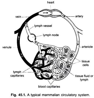
The blood vascular system is a closed system in vertebrates; it has a contractile heart and continuous tubes called vessels. The lymphatic system is an open system with lymph spaces. The blood vessels which carry blood away from the heart are arteries, which divide into thinner arterioles, branching into extremely thin and small capillaries.
The wall of a capillary is made of a single layer of tasselated endothelial cells. Every blood vessel, including the heart, has a lining of endothelial cells or endothelium. Blood does not come in contact directly with tissue cells.
Substances pass from and into capillaries through the tissue fluid contained in tissue spaces between cells. Exchange of substances between the blood and tissue cells of the body takes place through the capillary walls. This is brought about by pinocytosis in the endothelial cells of capillaries.
ADVERTISEMENTS:
Under an electron microscope the endothelial cells show many small vesicles which are invaginations of the plasma membranes. These vesicles move from one side of the cell and release their contents on the opposite side.
Thus, the blood gives up oxygen, nutrient material and hormones to the cells for metabolism, and the cells give out water, carbon dioxide, and nitrogenous wastes into the blood to carry them to the excretory organs for their quick elimination.
The capillaries form a network in all body tissues except cartilage and epithelium. From the capillaries the blood passes into thin venules which combine to form veins which carry blood towards the heart. But some veins (portal veins, renal veins, and hepatic vein) have capillaries which are just like those of arteries. But all blood does not pass through capillaries into venules.
There are also some through channels between arterioles and venules lying in some organs, such as the skin. There are also arteriovenous anastomoses between arterioles and venules found in the digits. The function of such connections is not clearly known, though it is claimed that they regulate blood pressure and circulation.
ADVERTISEMENTS:
At some places exchange of material between tissues and blood takes place through thin-walled spaces or sinusoids. In certain organs a blood vessel may form a coiled network of tiny blood vessels called rete mirabile (kidneys, air bladder).
Parts of Circulatory System:
In chordates, the circulatory system is of closed type. This type is also found in annelids (invertebrates). Another type is of open type in which capillaries are not found. It is found in molluscs and arthropods. Here the blood flows through arteries into various organs, which pass through blood spaces or sinuses and then again into vessels (veins) to the heart.
ADVERTISEMENTS:
The circulatory system includes the heart, arteries, veins, capillaries and blood. Heart is a modified blood vessel with muscular walls, which periodically contracts to pump the blood into the various parts of the body through definite vessels.
Arteries and their branches form an arterial system which carries the blood from the heart. Veins and their tributaries constitute a venous system which takes the blood from the capillaries of arteries or arterioles and carry that to the heart.
Portal System:
In the portal system, the blood is not returned directly to the heart, but there is an interposing organ (liver or kidney) in the course of the returning blood. The vein bringing blood starts in capillaries and ends in capillaries, the vein concerned acting both as afferent and efferent vessel, the afferent vessels end in capillaries just like arteries, then the blood is collected into systemic veins.
All vertebrates have a hepatic portal system in which blood passes into two sets of capillaries in the liver. Lower vertebrates and embryos of higher vertebrates have a renal portal system also in which blood passes through two sets of capillaries in the kidney before reaching the heart. The capillaries in the pituitary gland form a pituitary portal system which is a small but an important system.
Lymphatic System:
It is found in chordates except cyclostomes and cartilaginous fishes. It includes lymph and lymph vessels. Lymph is a tissue fluid found amongst the body cells. It is blood plasma minus red blood corpuscles and some proteins.
Lymph capillaries forming a network of thin blind-ending vessels, which collect lymph. Lymph vessels are thin-walled vessels formed by the union of blood capillaries. These empty into veins. Lymph nodes are found on lymph vessels in mammals. These form lymphocytes of blood used for body defence against diseases.
Evolution of Heart in Vertebrates:
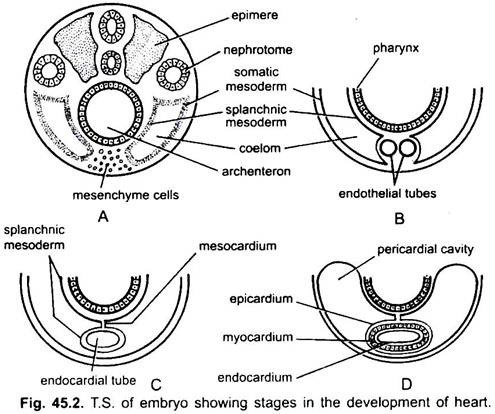
The heart is an unpaired organ but its origin is bilateral. In an embryo the mesenchyme forms a group of endocardial cells below the pharynx. These cells become arranged to form a pair of thin endothelial tubes. The two endothelial tubes soon fuse to form a single endocardial tube lying longitudinally below the pharynx.
ADVERTISEMENTS:
The splanchnic mesoderm lying below the endoderm gets folded longitudinally around the endocardial tube. This two-layered tube will form the heart in which the splanchnic mesoderm thickens to form a myocardium or muscular wall of the heart and an outer thin epicardium or visceral pericardium. The endocardial tube becomes the lining of the heart known as endocardium.
Folds of splanchnic mesoderm meet above to form a dorsal mesocardium which suspends the heart in the coelom. Soon a transverse septum is formed behind the heart which divides the coelom into two chambers, an anterior pericardial cavity enclosing the heart and a posterior abdominal cavity.
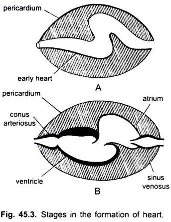
The heart is a straight tube but it increases in length and becomes S-shaped because its ends are fixed. Appearance of valves, constriction, partitions in the heart, and differential thickenings of its walls form three or four chambers in the heart.
1. Single-Chambered Heart:
In Amphioxus (primitive chordate), a true heart is not found. A part of ventral aorta beneath the pharynx is muscular and contractile and acts as heart.
2. Two-Chambered Heart:
In cyclostomes, there are four chambers arranged in a linear order- a thin-walled sinus venosus, a slightly muscular atrium (auricle), a muscular ventricle and a muscular conus arteriosus or bulbus cordis. It lies in the body cavity in which other visceral organs are also present.
Out of four chambers, only atrium and ventricle correspond to the four chambers (paired atria and paired ventricles) of the higher vertebrates. In the evolution of heart many changes have taken place.
Elasmobranchs:
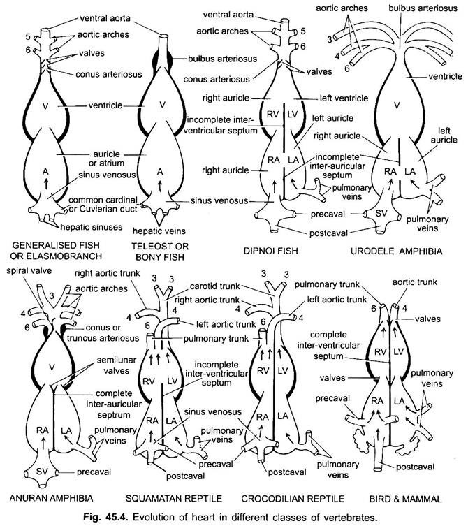
Except Dipnoi, the circulatory system in fishes from cyclostomes to teleosts, only unoxygenated blood goes to the heart, from there it is pumped to the gills, aerated and then distributed to the body. The heart of cartilaginous dogfish is muscular and dorsoventrally bent S-shaped tube with four compartments in a linear series.
They are sinus venosus and atrium for receiving venous blood, and a ventricle and conus arteriosus for pumping this blood. The heart is a branchial venous heart. The sinus venosus and conus arteriosus are accessory chambers. Atrium and ventricle are true chambers, thus, it is a 2-chambered heart.
The sinus venosus opens anteriorly into atrium through sinu-atrial aperture guarded by a pair of valves. Atrium lies dorsal to ventricle and opens ventrally into ventricle through an atrio-ventricular aperture guarded by a pair of valves. The thick-walled, muscular ventricle opens into a narrow conus arteriosus containing valves in two series.
The heart is enclosed within pericardial cavity separated from body cavity by a transverse septum. Conus pierces the pericardium and becomes continuous with the ventral aorta. Pericardial cavity communicates with the body cavity through two perforations in the transverse septum.
Teleosts:
Their heart resembles to that of clasmobranchs. In teleosts, the conus is reduced and has a single pair of valves. The proximal part of ventral aorta close to conus becomes greatly enlarged and thick-walled, called bulbus arteriosus. It is elastic and dilates at the time of ventricular contraction. The heart is, thus, 2-chambered with a single circulation of blood.
3. Three-Chambered Heart:
Dipnoi:
In diphoans a septum divides the atrium into a right and left chamber. This is correlated with the use of the swim-bladder as an organ of respiration and represents the first step toward the development of the double-type circulatory system whereby both oxygenated and unoxygenated blood enter the heart and are kept separate.
Blood from right auricle of the lungfish passes into the right ventricle and is then pumped into the primitive lung-like gas bladder by pulmonary arteries which branch off from the sixth pair of aortic arches. The oxygenated blood returns to the left atrium by way of pulmonary veins like amphibians.
Amphibia:
In amphibians, the dorsal atrium shifts anterior to ventricle. The sinus venosus opens into right atrium dorsally and not posteriorly. The atrium is completely divided into right and left chambers and has no foramen ovale in the inter-auricular septum, which remains open in dipnoans.
Deep pockets develop in the ventricular cavity. The conus arteriosus divides into systemic and pulmonary vessels by a spiral valve. In lung less salamanders, the inter-atrial septum is incomplete and pulmonary veins are absent.
Reptilia:
In reptiles, the heart is further advanced. The atrium is always completely separated into a right and left chamber, and in many forms the sinus venosus is incorporated into the wall of the right atrium. The ventricle is also partly divided by a septum in most reptiles, and in the alligators and crocodiles is completely two-chambered.
This means that oxygenated blood coming from the lungs to the left side of the heart is essentially separated from the non-oxygenated blood from the body to the right side. Thus, in crocodilians, the two types of blood is completely separated, and nearly complete in other reptiles, but some mixing does occur in other parts of the circulatory system.
The embryonic conus arteriosus splits into three instead of two vessels:
(i) Pulmonary arch carrying blood to the lungs from right side of the ventricle.
(ii) Right systemic aorta carrying blood from left side of the ventricle to the body by way of right fourth aortic arch.
(iii) Left systemic comes from the right ventricle to the left fourth aortic arch.
At the point of contact with the systemic aorta from the left ventricle, even in crocodilians, an opening between the two is present, called the foramen of Panizzae where there may be some mixing of the two types of blood. Thus, reptilian heart represents the transitional heart against amphibian heart-2 complete auricles and 2 incomplete ventricles with a little mixing of blood in right and left systemic.
4. Four-Chambered Heart:
Aves and Mammalia:
In birds, the ventricle is completely divided into two, so that the heart is four chambered (2 auricles and 2 ventricles). There is complete separation of venous and arterial blood. The systemic aorta leaves the left ventricle and carries blood to the head and body. While the pulmonary artery leaves the right ventricle and carries blood to the lungs for oxygenation.
Thus, there is double circulation in which there is no mixing of blood at any place. The sinus venosus is completely incorporated into right auricle, which receives two precavals and a postcaval. The left auricle receives oxygenated blood through pulmonary veins, conus arteriosus is absent, the pulmonary aorta arises from the right ventricle, and single systemic aorta arises from the left ventricle, and both have valves at their bases.
Modifications of Aortic Arches in Vertebrates:
Embryonic Arteries:
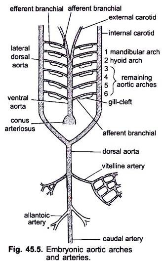
When the heart is being formed in a vertebrate embryo, a blood vessel called ventral aorta appears mid-ventrally below the pharynx, which soon gets connected to the conus arteriosus. The ventral aorta arising from the heart runs forward beneath the pharynx and divides anteriorly into a pair of external carotid arteries into head.
The ventral aorta gives off 6 pairs of lateral aortic arches at equidistance which run through the visceral arches. Each aortic arch has a ventral afferent branchial artery carrying venous blood to the gill and a dorsal efferent branchial artery taking oxygeneted blood from the gill. The efferent branchial arteries of either side join dorsally with the lateral dorsal aorta or radix aorta which enters into the head as internal carotid artery.
The first aortic arch is a mandibular aortic arch, the second is a hyoid aortic arch, the remaining ones are called third, fourth, fifth and sixth aortic arches. The lateral dossal aorta fuse behind the pharynx to form a dorsal aorta which is continued mid-dorsally into the tail as a caudal artery.
From the dorsal aorta paired and unpaired arteries arise which supply various organs of the body. In an embryo with a yolk sac, a pair of vitelline arteries arises from the dorsal aorta and supplies the yolk sac. In embryos of amniotes a pair of umbilical or allantoic arteries arises from the dorsal aorta supplying blood to the allantois.
In an adult the vitelline arteries fuse to form the main mesenteric artery, the major part of the allantoic arteries is lost, but their remnants form hypogastric or internal iliac arteries.
Aortic Arches in Vertebrates:
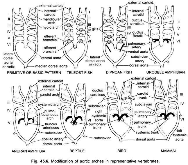
In various adult vertebrates, the arterial system appears to be different, but they are built on the same basic fundamental plan. The difference is due to increasing complexity of heart due to a change in respiration from gills to lungs. There is a progressive reduction in the number of aortic arches in the vertebrate series.
Cyclostomata:
In Petromyzon, there are 7 pairs of aortic arches. In other cyclostomes these vary from 6 pairs in Myxine and 15 pairs in Eptatretus.
Pisces:
Although six is considered to have been the basic number of aortic arches for fishes. This number is reduced to five even in sharks and rays with the loss of the first pair, the mandibular aortic arch or it is represented by an efferent pseudobranchial artery. In most bony fishes both the mandibular (i) and hyoid (ii) aortic arches disappear or are much reduced.
In Polypterus and Dipnoi (lung-fishes), gills are not well developed. So the pulmonary artery arises from the efferent part of 6th arch on either side which supplies blood to the air bladder or lung. In elasmobranchs and Dipnoi each arch has one afferent and two efferent branchial arteries (formed by splitting) in each gill. In bony fishes each gill has one afferent and one efferent artery.
In tetrapoda aortic arches do not break up into afferent and efferent parts because true internal gills are absent. In all tetrapoda the first and second arches disappear.
Amphibia:
Here the aortic arches show modification due to loss of gills and appearance of the lungs. In urodeles there are external gills present as respiratory organs in addition to lungs. The III, IV, V and VI aortic arches are present, though the fifth pair is much reduced in Siren, Amphiuma and Necturus. The aortic arches are not broken in the external gills into afferent and efferent portions, because branches arising from IV, V, and VI aortic arches form capillaries in the external gills.
The lateral dorsal aortae between the III and IV aortic arches persist as a vascular connection, the ductus caroticus. The VI aortic arch forms the pulmo-cutaneous arch or artery on either side taking blood to the lung and skin. It also retains a connection with the lateral dorsal aorta known as a ductus arteriosus (duct of Botalli).
In the larva of anuran (frog tadpole), arrangement of aortic arches are like an adult urodele due to presence of gills. At metamorphosis, with the loss of gills, I, II, and V aortic arches disappear completely, only the IIIrd, IVth and Vlth aortic arches are present. The lateral dorsal aorta between the third and fourth aortic arches (ductus caroticus) also disappears. Thus, the third aortic arch along with a part of the ventral aorta becomes the carotid arch carrying oxygenated blood to the head region.
The fourth aortic arch along its lateral dorsal aorta forms the systemic arch. The sixth aortic arch becomes the pulmocutaneous arch supplying venous blood to lungs and skin. The ductus arteriosus disappears during metamorphosis. Thus, adult anurans have only III, IV and VI aortic arches. These are also retained by amniotes.
Reptilia:
In reptiles, the gills are fully replaced by lungs. Only III, IV and VI aortic arches are present. With the partial separation of the ventricle into two parts, the distal portion of the conus arteriosus and the entire ventral aorta are split into three vessels, i.e., two aortic or systemic and one pulmonary. Right systemic arch (IV) arise from the left ventricle carrying oxygenated blood to the carotid arch (III). The left systemic (IV) and pulmonary aortae (VI) take their origin from the right ventricle. The left systemic carries deoxygenated or mixed blood to the body through dorsal aorta.
While the pulmonary artery takes deoxygenated blood to the lungs. The ductus caroticus disappears, but it persists in snakes and some lizards (Uromastix). The ductus arteriosus disappears in most reptiles though it persists in a reduced form in Sphenodon and some turtles. Due to mixing of blood, reptiles are cold blooded animals like fishes and amphibians.
Aves:
In birds, the III, IV and VI aortic arches are present. They follow the general pattern of reptiles with some differences. With the complete division of the ventricle into two parts, the conus arteriosus and ventral aorta have split to form two vessels, systemic aorta arising from the left ventricle and a pulmonary aorta from the right ventricle.
Third aortic arch with remnants of lateral and ventral aortae forms the carotids which arise from systemic aorta. Fourth aortic arch forms the systemic aorta on the right side only. It unites with the lateral aorta of its own side and forms the dorsal aorta. Part of the fourth aortic arch of the left side forms the left subclavian artery, the rest along with its lateral dorsal aorta disappears. The sixth aortic arch forms the pulmonary aorta. Ductus caroticus and ductus arteriosus disappear.
Mammalia:
In mammals also the III, IV and VI aoric arches persist. The ventricle is divided completely into two parts.
The conus arteriosus and ventral aorta split to form two vessels:
(i) A systemic aorta arising from the left ventricle, and
(ii) A pulmonary aorta from the right ventricle. Third aortic arch with remnants of lateral and ventral aortae forms the carotid arch.
Fourth aortic arch forms the systemic aorta on the left side only, while on the right side its proximal portion forms an innominate and right subclavian artery, the rest along with its lateral dorsal aorta disappears. Sixth aortic arch forms the pulmonary aorta. The ductus arteriosus degenerates but it persists in some until hatching or birth in a reduced form on the left side as a thin ligamentum arteriosum.
Venous System:
Embryonic Veins:
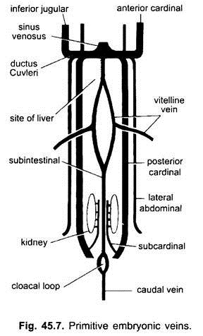
In all the vertebrate embryos, the venous system is simple and similar. The veins are mostly paired and symmetrically arranged. In embryos without a yolk sac, a sub- intestinal vein is formed in the splanchnic mesoderm below the gut.
It loops around the anus and is continued posteriorly as:
(i) A caudal vein into the tail. In all embryos having a yolk sac (whether containing yolk or not), a pair of vitelline veins arises from the yolk sac and joins the posterior part of the developing heart which becomes the sinus venosus. In fact the fusion of these vitelline veins is responsible for the heart formation in bony fishes, reptiles and birds. Each vitelline vein at its posterior end joins the subintestinal vein formed in the same way as in the embryos having no yolk sac.
(ii) A pair of subcardinal veins arises between the kidneys and joins the caudal vein.
(iii) Paired anterior and posterior cardinal veins are formed, which carry the blood from the head and posterior parts of the body respectively. The anterior and posterior cardinal veins of each side unite to form a ductus Cuvieri or common cardinal vein which passes inwards through the transverse septum to enter the sinus venosus.
In fishes and salamanders (urodeles) an inferior jugular vein comes from the ventral side of the head to join the common cardinal vein. It has no homologue in other vertebrates. In an amniotes a pair of lateral or ventral abdominal veins comes from the body wall to enter the common cardinal veins.
Pisces:
Common cardinal vein (duct of Cuvier) enters the sinus venosus from each side and is formed by the fusion of anterior and posterior cardinals. Blood from the head is collected by the anterior cardinals, and blood from the kidneys and gonads is collected by the posterior cardinals.
Also entering the ducts of Cuvier are the paired lateral abdominal veins which receive blood from the body wall and paired appendages. The renal portal system consists of the caudal vein and two renal portal veins situated lateral to the kidneys which capillarised in the kidneys.
Hepatic portal system carries blood from the stomach and intestine and returns it to the liver, where, after passing through series of sinusoids, it enters the sinus venosus by way of paired hapatic veins.
In teleosts, the lateral abdominal veins are lacking so that blood from the subclavians, draining the pectoral appendages, enters the sinus venosus directly, and the blood from iliac veins, draining the pelvic appendages, passes into the postcardinals.
In dipnoans, a single ventral abdominal vein is present, presumably derived from a fusion of lateral abdominals. This vein receives blood from the ilacs by way of paired pelvic veins and enters the right duct of Cuvier. Furthermore, from the right postcardinal system, a new vein, the postclaval makes its appearance that is of major importance in higher vertebrates. It is connected with the caudal vein and passes forward through the liver to the sinus.
In embryos of amniotes the lateral abdominal veins are known as umbilical or allantoic veins because they drain the allantois. The vitelline and umbilical veins are lost at birth, only their remnants persist.
Modifications of Veins:
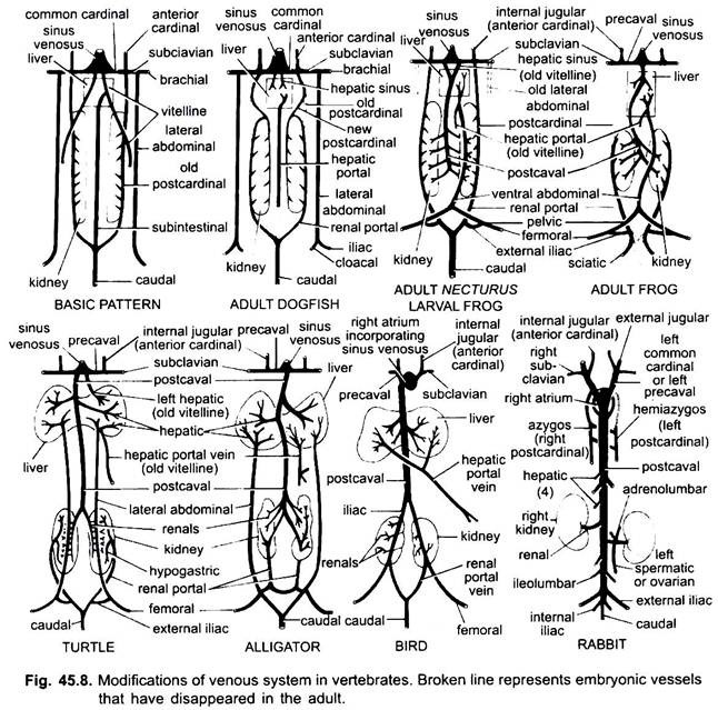
As in the case of arteries, the veins of different vertebrates are arranged on the same fundamental plan. Any variations shown by them follow a sequence in the vertebrate series. In its development the venous system of higher vertebrates passes through the stages seen in embryos of lower forms.
1. When the liver is being formed the proximal part of the vitelline veins or subintestinal veins forms the hepatic veins between the liver and the heart, the distal part of the left vitelline vein or subintestinal vein becomes the hepatic portal vein which forms sinusoids in the liver to give rise to a hepatic portal system present in all.
2. Anterior cardinal veins persist as internal jugular veins.
3. Except in fishes, the common cardinal veins become the precaval veins which enter the sinus venosus (amphibians, reptiles) or the right auricle (birds, mammals). A subclavian vein is formed in each forelimb which joins the precaval vein.
4. The caudal vein loses its connections with the subintestinal and subcardinal veins, its anterior part splits into two branches which join the posterior cardinal veins in all except mammals.
5. Posterior cardinal veins persist as such in fishes, but in others each breaks up into two portions, an anterior portion which disappears in amphibians, reptiles, and birds, but in mammals the right anterior portion forms an azygos vein, the left one may form a hemiazygos vein which loses its connection with the precaval and joins the azygos vein by a transverse anastomosis; the posterior portions of the posterior cardinal veins which have joined the caudal vein, become the renal portal veins.
The renal portal vein of each side forms capillaries in the kidney to form a renal portal system which is complete in fishes, amphibians, and reptiles, but becomes much reduced in birds, and is absent in mammals because the posterior portions of posterior cardinal veins disappear in mammals.
6. The renal portal veins of fishes collect blood only from the tail, but in amphibians, reptiles, and birds they also usurp the veins from the legs.
7. In some fishes and all tetrapoda the vitelline veins join the subcardinal vein to form a postcaval vein. In amphibians and reptiles the postcaval vein extends to the posterior ends of the kidneys, in birds it joins the renal portal veins, thus, reducing the renal portal system, in mammals the postcaval vein joins the veins from the legs and tail, so that the renal portal system is completely eliminated.
8. The two lateral abdominal veins persist as such in fishes, but from Dipnoi upwards they fuse to form an anterior abdominal vein which joins the hepatic portal vein near the liver, the anterior abdominal vein, thus, connects the renal portal and hepatic portal systems. An iliac vein is formed in each hindlimb which joins the anterior abdominal vein. The anterior abdominal vein begins to lose its importance in reptiles, in birds it is modified to form the epigastric and coccygeo-mesenteric veins, in mammals it disappears, except in Tachyglossus.
9. In air-breathing forms, pulmonary veins grow out from the left auricle and enter the lungs.
10. The umbilical (allantoic) veins of the embryo disappear when the lungs become functional as respiratory organs.
Tetrapoda:
The venous system of amphibians very much resembles that of lungfishes except that the abdominal vein enters the hepatic portal system rather than the sinus venosus. Anterior cardinals persist as internal jugular veins in all adult tetrapods. Inferior jugular veins are absent. Common cardinals become the anterior vena cavae or precavals, which join sinus venosus in amphibians and reptiles.
In birds and mammals, precavals directly enter the right auricle of heart, because sinus venosus is absent. In man, cat, etc., left precaval is lost so the blood of left side enters the right precaval through a brachio-cephalic branch. In lungless salamanders, the pulmonary veins are absent since inter-atrial septum is incomplete.
The post cardinal veins join the caudal vein posteriorly and precavals anteriorly to form the common cardinals in larval frog and Necturus.
In reptiles, there is greater development of pulmonary veins and postcaval vein and reduction in the importance of renal portal system which brings blood to the kidneys from the posterior part of the body.
In birds, there are two functional precaval veins formed by the union of the jugular and subclavian on each side, and a complete post caval. It receives blood from the limbs by way of the renal portals, which pass through kidneys, but do not break up into capillaries and are not comparable to the renal portals of lower vertebrates.
In mammals, there may be one or two precaval veins. There is a single postcaval vein, whose embryological development is very complicated. All the caval veins enter the right auricle directly, as the sinus venosus is absorbed into the wall of this chamber of the heart in embryonic life. There is no renal portal system although the hepatic portal system very much resembles that of other vertebrates.
