Both layers of integument have given rise to various types of derivatives. The epidermis gives rise to integumentary glands, epidermal scales, horns, digital structures, different corneal structures, feathers, and hairs. The dermis forms dermal scales of fishes and of some reptiles, plates or scutes in reptiles, fin rays in fishes and antlers in mammals.
I. Epidermal Derivatives:
Epidermal derivatives are epidermal glands (unicellular and multicellular), epidermal scales and scutes, horns, digital structures (claws, nails and hoofs), feathers and hairs.
1. Epidermal Glands:
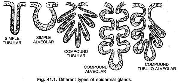
Epidermal glands are formed from the Malpighian layer of the epidermis. They arise from the epidermis and often penetrate the dermis. According to their structure they are unicellular or multicellular, tubular or alveolar and simple or compound (branched) glands. These are lined by cuboidal or columnar cells.
ADVERTISEMENTS:
(a) Unicellular glands are single modified cells found among other epithelial cells, they are present in amphioxus, cyclostomes, fishes and larvae of amphibians. Unicellular glands are known as mucous cells or goblet cells. They secrete a protein mucin which combines with water to form mucus which lubricates the surface of the body. Other unicellular glands are granular cells and large beaker cells of cyclostomes and fishes, they also secrete mucus.
(b) Multicellular glands are of two types:
(i) Tubular glands are multicellular tubes of uniform diameter formed as ingrowths of the Malpighian layer into the dermis, e.g., glands of Moll on the margin of the human eyelids. Tubular glands may become coiled at the base deep in the dermis, e.g., sweat or sudoriferous glands of mammals, or they may divide into many tubules which are then called compound tubular glands, e.g., mammary glands of females and of males in monotremes and primates, etc., and gastric glands in stomach.
(ii) Alveolar or saccular glands are multicellular down growths of the Malpighian layer into the dermis, having a tubular duct whose terminal parts form a rounded expansion to become flask-shaped, e.g., mucous and poison glands of amphibians. Alveolar glands may branch into many lobules which finally open into a common duct, they are then called compound alveolar glands, e.g., mammary glands of eutherians, and salivary glands.
Kinds of Epidermal Glands:
According to function, the epidermal glands of vertebrates are of the following types:
i. Mucous Glands:
They may be unicellular or multicellular. The unicellular glands are mucous gland cells, granular cells and beaker cells of amphioxus, cyclostomes and fishes. They secrete mucus which keeps the skin moist and slippery, and also affords protection against bacteria and fungi. Mucous cells and granular cells lie near the surface, but the beaker cells lie more deeply and extend from the Malpighian layer to the surface.
ADVERTISEMENTS:
Multicelluar mucous glands are alveolar found in some fishes and amphibians. They occur all over the surface of the body and produce mucus for lubricating the skin and in amphibians they keep the skin moist to aid in respiration.
ii. Poison Glands:
Amphibians also have alveolar poison glands which are larger but less numerous than mucous glands. In toads masses of poison glands form parotoid glands behind the head. The secretion of poison glands has a burning taste and is used as a defence. Caecilians have giant poison glands.
Some tubular glands are found on the feet and suctorial discs of tree frogs which aid in climbing. Tubular glands are also found on the swollen glandular thumb parts of male frogs and toads during the breeding season. They aid in clasping the female during amplexus.
iii. Luminescent Organs or Photophores:
They are found in longitudinal rows near the ventral side of the body in those fishes which live in deep sea where no light penetrates. Each photophore is a group of epidermal cells lying in the dermis.
Each photophore has a lower layer of luminous cells below which is a layer of reflecting pigment cells, and the upper layer of mucous cells forms a lens. The glandular cells produce phosphorescent light which is transmitted to the outside by other cells. The light helps to attract the prey of deep sea fishes.
iv. Femoral Glands:
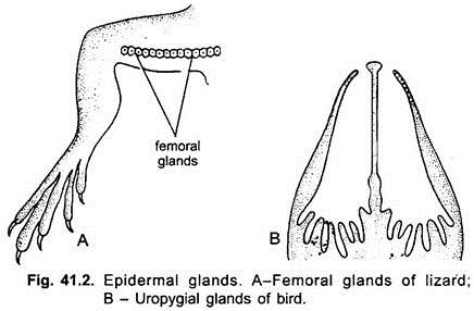
Femoral glands are found in male lizards (e.g., Uromastix) below the thighs in a row from the knee to the cloaca. They secrete a sticky substance which hardens into short spines that are used for holding the female during copulation.
ADVERTISEMENTS:
v. Uropygial Glands:
These are the only glands in birds, and they are best developed in aquatic birds. Uropygial glands are branched alveolar glands located on the dorsal side at the base of the tail or uropygium in the form of swelling. They secrete an oil which is odoriferous and attracts the opposite sex. The oil contains pomatum which is picked up with the beak and used for preening and waterproofing the feathers.
vi. Sweat Glands:
The largest number and variety of epidermal glands are found in the skin of mammals. They are tubular or alveolar and multicellular. Sudorific or sweat glands are long, coiled tubular glands embedded deep in the dermis. Their upper part forms a duct which opens on the surface by a pore and the lower coiled part lies in the dermis surrounded by a network of blood capillaries.
Sweat is secreted by sweat glands continuously, may or may not be visible. Sweat contains a large amount of water having dissolved salts of sodium, potassium and urea. Sweat removes some metabolic wastes and regulates the body temperature by evaporation.
Sweat glands are not uniformly distributed. In man they are more numerous on palms, soles, and arm pits. In cats, rats, and mice they are confined to the soles of the feet. In rabbit they are around the lips; in bats on the sides of head; in ruminants on the muzzle and the skin between the digits and in hippopotamus they are found only on the pinnae.
There are no sweat glands in Tachyglossus, Sirenia, and Cetacea. In some mammals the secretion of sudorific glands is red- coloured. In hippopotamus it is red and spreads on the head and back. The male giant kangaroo Macropus rufus secretes red sweat. Modified sudorific glands form glands of Moll in the margins of the human eye in connection with eye-lashes.
Ceruminous glands in the external ear passages of mammals are modified sweat glands and secrete a waxy substance which combines with the secretion of sebaceous glands to form earwax which catches dust. Oil glands form ceruminous glands in the external ears of some gallinaceous birds.
vii. Sebaceous Glands:
Sebaceous glands are alveolar glands opening in hair follicles containing hairs. They also independently open at the skin surface around the genital organs, tip of the nose, and edges of the lips. Sebaceous glands secrete an oil (sebum) to lubricate the hairs and also cover the skin with a film of oily coating. The oily secretion of sebaceous glands contains waxes, fatty acids, and cholesterol, which makes the skin pliable.
Sebaceous glands are absent in Manis (pangolin), and Sirenia, and Cetacea which practically have no hairs. Modified sebaceous glands form Meibomian glands in the eyelids, each has a long straight duct into which separate alveoli open.
They produce an oily secretion which forms a film over the lacrimal fluid or tears holding them evenly on the surface of the eyeball for keeping the eye moist, in weeping the oily film are broken and tears flow out. Ceruminous glands of external auditory meatus are modified sebaceous glands. Their greasy or waxy secretion, called the cerumen traps the insects and dust particles.
viii. Scent Glands:
Scent glands are modified sebaceous glands or sweat glands. Their secretion is an allurement to the opposite sex. Scent glands are located in the deer family on the head near the eyes. Skunks and carnivores have scent glands around the anus, and pigs and goats have scent glands between their toes.
ix. Preputial Glands:
These are found in many kinds of mammals. In beaver, large sacs containing a secretion known as castoreum lie beneath the skin on either side of penis and open by ducts into the prepuce.
x. Mammary Glands:
These are characteristic of mammals. They secrete milk generally in the females for nourishment of the young. In monotremes both sexes may secrete milk, this condition is called gynaecomastism which is not a normal condition. Mammary glands of monotremes are compound tubular glands, while in other mammals they are compound alveolar.
Mammary glands of monotremes have no nipples, they open into pits on the surface of the skin, and the young ones obtain milk by licking tufts of hairs. In others the mammary glands open by their ducts separately into a nipple.
The nipple is a raised outgrowth of the breast, bearing opening of mammary glands (true teats). The false teat has an elongated cistern into which the mammary glands open by their ducts. From the cistern a tube leads to the surface of the nipple.
Mammary glands along with fat form integumentary swellings called mammae or breasts. The number and location of mammae varies in different mammals. The number ranges from two in many mammals to 25 in opossum. Mammae may run along two ventral milk lines from the arm pits to the groin, or they may be axillary, thoracic, abdominal or inguinal in position.
2. Epidermal Scales and Scutes:
Epidermal Scales:
After the evolutionary loss of dermal scales of fishes, amniotes developed an entirely new type of scale derived from the epidermis. The skin of vertebrates is rarely naked, and it is usually provided with protective scales, bony plates, feathers or hairs. There are no epidermal scales in fishes and amphibians. They appear for the first time in reptiles.
They are cornified derivatives of the Malpighian layer and are generally shed and replaced. The scleroprotein called the keratin is continually accumulated in the permanently growing layer of the epidermis called the stratum germinativum.
These cells are called cornified and they become dead. Thus, the stratum corneum cells become cornified and form hard horny structures, such as scales and scutes in reptiles, beaks, claws, horns, nails, hoofs, feathers and hairs in different vertebrates.
Scales are most noticeable on lizards and snakes. They are continually being produced by the permanently growing layer of the stratum germinativum and are generally folded so as to overlap one another. When they are fully grown they become separated from the stratum germinativum and appear as non-living, cornified structures.
The epidermal scales form a protective covering of the body (a continuous armour). The scales in snakes and lizards are continuous and undergo ecdysis periodically. Before ecdysis, the new scales that will replace the old ones are formed. The entire corneal layer of scales is shed as whole in snakes. The old epidermal covering becomes loosened first in the head region.
This skin, including even the transparent plates over the eyes, is turned back and the snake finally crawls out of the old covering, leaving the old covering inside out. The scales on the ventral side in most snakes differ from their other scales in being long and transversely arranged, they aid in locomotion.
Turtles and crocodiles have different kinds of scales which do not overlap, nor undergo periodic ecdysis, but the scales are gradually worn off and replaced. Large epidermal scales, such as those on the shell of turtles and on the head of snakes, are generally called scutes. In birds the scales are confined to the shanks and feet and some at the base of the beak.
They generally overlap as in snakes and lizards. In mammals epidermal scales are found on the tail and paws of rats, mice, beavers, musk rats and shrews. These scales are not much cornified, nor do they undergo ecdysis; hairs project from beneath the scales. In scaly ant-eaters large epidermal scales cover the entire body, except the ventral side, the scales are reptilian and undergo ecdysis singly.
In armadillos there are large scales which fuse to form plates on the head, shoulders and hips; in the middle of the body, except mid-ventrally, the scales fuse to form ring-like bands; these scales do not undergo ecdysis but are gradually worn off and replaced.
Some epidermal scales of the tail of a rattle-snake are modified to form a rattle. It consists of a series of old dried scales. Rattles of rattle snakes represent horny remnants of the skin which adhere to the base of the tail and are not lost during ecdysis.
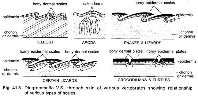
During ecdysis the scale at the tip of the tail is not shed and it forms a ring. Thus, after several ecdysis a series of rings form a rattle, each new ring being larger than its predecessor. In older rattle snakes the terminal rattles are often lost. In tortoises and turtles, the dermal plates forming the carapace and plastron are covered externally with cornified epidermal scales.
These are not shed regularly like lizards and snakes. A few turtles lack scales and have a leathery skin. The bodies of alligators and their relatives are also covered with epidermal scales which gradually wear off and are replaced.
In turtles, tortoises and modern birds there are no teeth. Each jaw bone being covered with horny sheath formed of several fused plates or scales which form a beak. In monotremes there is a soft bill which differs from the beak of birds is not being covered with modified epidermal scales.
3. Horns:
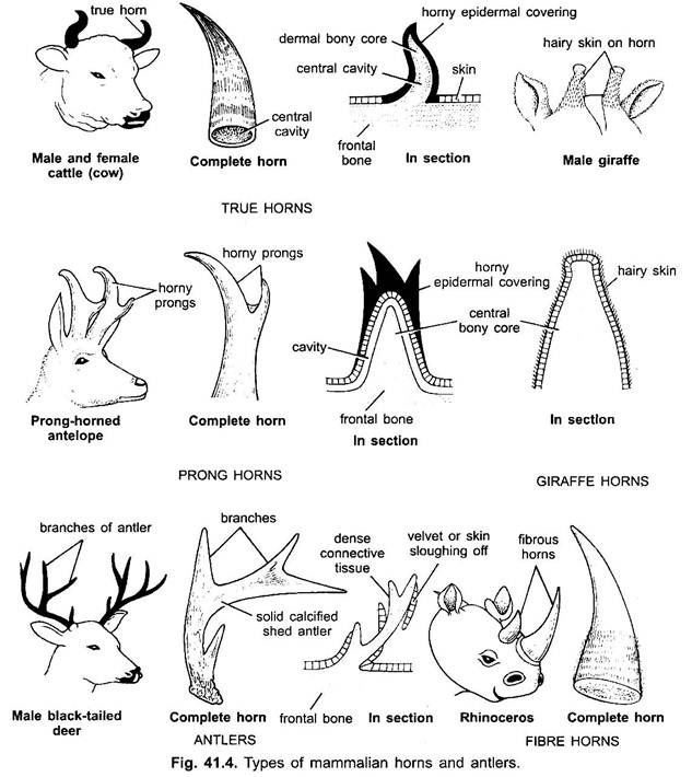
Horns are found in ungulate (even-toed hoofed) mammals only. True horns of the hollow type are found in pronghorn, cattle, antelope, sheep and goats consist of an inner core of bone which is an outgrowth from the frontal bone. It is encased in a keratinised, epidermal covering. True horns continuously grow throughout life and are not shed.
Following types of horns are recognised:
(a) Rhinoceros or keratin fibre horn has no skeletal element. It is made by keratinised cells of the epidermis and consists of matted keratin fibres bound together, but its fibres are not true hair. It is a permanent epidermal structure and if broken it grows again. There is one horn in the Indian rhinoceros and two in the African species.
(b) Pronghorn is a true horn, consists of a permanent projection of the frontal bone covered by a hard, horny epidermal sheath. The sheath is forked bearing one to three prongs made only of horny sheath. The horny sheath is shed annually and is replaced by another which grows from the skin that surrounds the core. It is found in Russian antelope Antilocapra.
(c) Giraffe Horns:
They develop from cartilaginous protrusions which are present at birth. They ossify and fuse at the top of the skull, where they appear as knobs permanently covered with living skin and hair. Giraffe possesses three of these knobs, one is median and anterior to the other two. These horns are short, unbranched and are permanent, and are present in both sexes.
(d) Antlers are found in the males of deer family, but they are present in both sexes in reindeer and caribou. An antler consists of a branching solid outgrowth of the frontal bone formed of dense connective tissue. It is covered during growth by hairy, vascular skin called ‘velvet’. The velvet is shed exposing the antler naked when the antler reaches full growth.
Thus, the antler consists only of dermal bone. The bony antler is also shed annually after the breeding season, and a new antler develops. Antlers are solid mesodermal bone, but they are formed under the influence of the integument. Formation of antlers is controlled by the hormones of testes and anterior lobe of the pituitary.
4. Digital Structures:
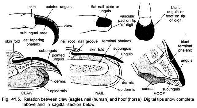
In amniota the distal ends of digits have claws, nails or hoofs formed from the horny layer of the epidermis. They grow parallel to the surface of the skin and are built on the same plan.
i. Claws:
Claws made their appearance first in the reptiles. A claw is made of a hard horny dorsal scale-like plate called unguis and a relatively soft ventral subunguis, both converge terminally and cover the terminal part of the last phalanx. Claws of reptiles and birds are similar but in mammals the subunguis is much reduced and is continuous with a pad at the end of a digit. In cat family claws are retractile.
ii. Nails:
They are found in primates. The dorsal unguis is large and flat and subunguis is soft and much reduced. Tip of the digit forms a sensitive and vascular pad over which the nail groove is present. It is formed by the invagination of epidermis. Growth of the unguis takes place from the nail root lying below the skin in the nail groove.
iii. Hoofs:
They are found in ungulates. The horny unguis is thick and around the end of the digit, and encloses the thickened subunguis which touches the ground. Subunguis surrounds the soft, horny cuneus. Tip of digit, thus, forms a pad containing a blunt phalanx. Nails and hoofs of mammals are modified claws. Whalebone plates of toothless whales are also the modification of stratum corneum.
5. Feathers:
Feathers are found only in birds and are formed from the epidermis in which the stratum corneum is highly specialised. Feathers are light, strong, elastic, waterproof and show many colours due to pigments and structural arrangement. The pigments are carotenoids and melanins. Carotenoids are frequently called lipochromes which are soluble in fat solvents like methanol, ether or carbon disulphide, and insoluble in water.
Animal red is zooerythrin and animal yellow is zooxanthin are the two groups of carotenoids. Melanins are soluble only in acids. Eumelanin granules vary from black to dark brown and phaeomelanin granules may be almost colourless to reddish brown.
Many of the feather colours are the product of both carotenoids and melanin. They form a protective covering (conserve body heat), provide buoyancy to the water birds during swimming and at rest, regulate body temperature and support the body in flight.
There are three kinds of feathers in birds, they are contour feathers, down feathers, and filoplumes. The development of feather is like that of scales.
Shedding and replacement of feathers is moulting which takes place gradually, one moulting usually takes an average time of six weeks. At the base of each feather follicle a dermal papilla persists from which new feathers will form, so that there is a continuous replacement of feathers throughout life. The replacement in some birds is seasonal, while in others it is gradual throughout the year.
Feathers of birds show varied and often brilliant colourations.
The colours are due to three factors:
(a) Pigment is deposited in the feathers during development, the colour that is seen is due to absorption of some wavelengths of light by the pigment, thus, black, red, brown, yellow, and orange colours are seen. Blue, most greens and violets are not the result of pigment but depend entirely on the feather structure. Blue feathers in blue birds do not contain any blue pigment, but they absorb all, but the blue rays of the spectrum are reflected. White colour is not due to white pigment, but is caused, due to reflection of light from the feather without absorption of any wavelengths of light rays;
(b) Structural arrangement or striations of feather surface are prismatic, these cause iridescence due to reflection of light, thus, producing iridescent hues, metallic colours, gray, and some shades of blue;
(c) A combination of pigments and prismatic striations of the feather produce green in which the yellow pigment combines with the structural blue. The colours of birds are for concealment, recognition, and sexual stimulation. The colour of various kinds of birds is under genetic control but it may be modified by internal and external factors.
The birds kept in captivity for several years get their plumage colour changed from red to yellow. In captive birds this change has been attributed by many to diet. Hormones also play an important role in the control of feather colour. Estrogenic hormone given to male before moult result in the assumption of female plumage. Similarly females may acquire male plumage by giving the testosterone.
Oxidation and abrasion are external factors that change in colour to a lesser or greater degree in most species of birds. Carotenoids fades in sunlight.
6. Hair:
Hair is found only in mammals. It projects at an acute angle from the skin. Hair covers the entire integument in most (furred mammals), but in others only traces are left, such as whales have only a few core hairs on the snout.
But during development the body of the embryos of all mammals is covered with a coating of fine hair called lanugo which is usually shed before birth and replaced by a new one. Hair is entirely epidermal in origin.
The hairs trap air which does not transmit body heat and, thus, act as insulators for the body. In some animals the colours of hair are protective. Hair in nostrils and ears prevent entry of dust, eyebrows and eyelashes protect the eyes, vibrissae are delicate organs of touch (act as tactile organs), hairs on the tail are used to drive away insects. In some animals, such as lions and some monkeys the mane distinguishes the male. Hairs are also modified in spines, scales, horns, etc., in some mammals.
Structure:
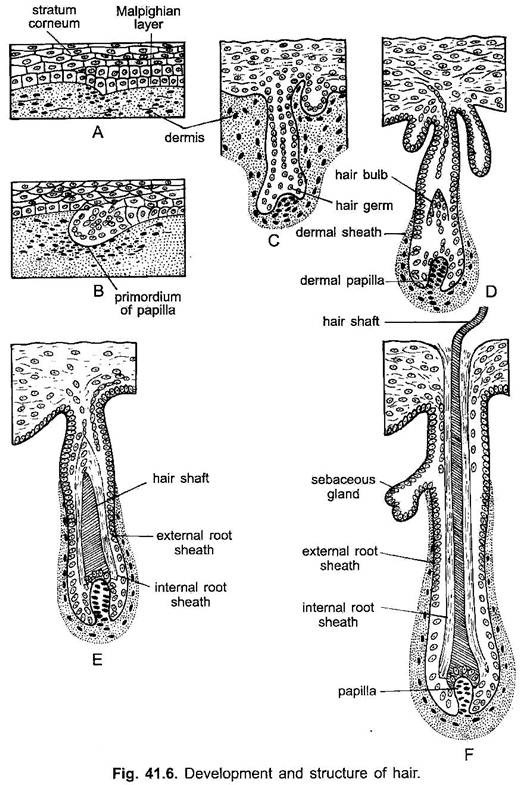
Hairs are not modified scales but are new outgrowths of the epidermis only. A hair has an upper projecting shaft and a lower root lying in a hair follicle which is a sunken pit in the dermis. The shaft is made of only dead, keratinised cells. The part of the hair protruding above the skin is dead.
At the base of the follicle the root is expanded into a bulb and growth of the hair takes place only in the root where the cells of the Malpighian layer divided actively. Below the bulb is a dermal papilla having connective tissue and blood vessels, it nourishes the hair. Beyond the bulb the cells gradually die so that the shaft is made of dead cornified cells.
The hair shaft has an external cuticle of transparent overlapping cells which have lost their nuclei, inside the cuticle is a cortex (middle part of hair) containing shrivelled cells and pigments, and a central core or medulla having air spaces. In the follicle the hair root is surrounded by two layers of hair sheath cells forming an outer and inner root sheaths. They do not extend beyond the follicle. A sebaceous gland opens into the upper part of the hair follicle for oiling the hair.
An arrector pili muscle composed of smooth fibres extends from the upper part of the dermis to the basal part of the hair follicle on the side towards which the hair slopes. It pulls the hair base causing the hair to stand when an animal is confronted with danger. Hair does not project vertically but at an acute angle from the skin.
Development of Hair:
A thickening of the epidermis (stratum germinativum) pushes into the dermis and becomes cup-shaped at its lower end. The dermis extends into the cup forming a hair papilla which has blood vessels for supplying nourishment. The epidermal down growth which at first is a solid cord of cells now splits to form a central shaft of cornified keratinised cells, and a space around it.
The epidermal cells around the space form the hair follicle. The lower part of the hair follicle becomes swollen and is known as a bulb. The cells of the follicle thickened and bud off a sebaceous gland. The central shaft by addition of new keratinised cells from the root grows in length and pushes through the solid epidermal cells to emerge outside the skin.
Thus, the development of hair differs from that of a feather. It is formed entirely from the solid column of epidermis, while in the feather there is a mesodermal feather pulp extending into the hollow quill.
II. Dermal Derivatives:
The scales arise from the dermis and are, thus, mesodermal in origin, they are found only in fishes, some reptiles and a few mammals. Ostracoderm fishes, the earliest known vertebrates, had an armour of large bony plates. These bony plates became very small in placoderms to give rise to cosmoid scales which are not found in any living forms today (except in Latimeria).
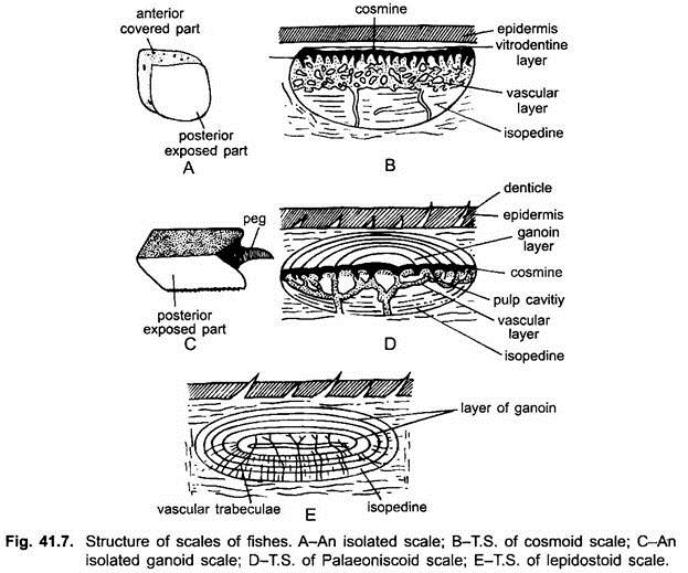
1. Cosmoid scales were also present in primitive Choanichthyes. A cosmoid scale had four distinct layers, the lowest layer is of isopedine or dentine resembled compact bone, the next layer was a spongy bone with vascular spaces consisting of pulp cavities having odontoblasts, the third layer was of hard compact cosmine with canaliculi and the outermost layer was thin but hard vitrodentine or enamel.
2. Ganoid scales are the other type of scales found in earliest primitive bony fishes are ganoid scales. This implies that two different lines of evolution with regard to scales appeared very early in the history of fishes. A ganoid scale has a basal layer of isopedine, above which there may be a reduced cosmine layer or it may be absent and the uppermost layer is made of a hard, translucent substance called ganoin. Ganoid scales with a reduced cosmine layer are found in Polypterus, and with no cosmine in Lepidosteus.
3. The evolution of the ganoid scale by the loss of its upper layer of ganoin gave rise to a thinner leptoid scale. There are two types of leptoid scales, namely, (i) cycloid and (ii) ctenoid scales. In the other line of evolution the cosmoid scale lost its three lower layers, only the fourth enamel-like dentine layer was retained and somewhat elaborated to form the placoid scale. Thus, in the present fishes there are four types of dermal scales- placoid, ganoid, ctenoid and cycloid scales.
Embedded in the dermis of elasmobranchs in oblique rows and projecting from the epidermis are dermal denticles of placoid scales forming an exoskeleton. The placoid scales were evolved by loss of some layers from the cosmoid scales. Placoid scales are found only in elasmobranchs, in sharks they are very small and give the skin a rough texture, but in skates they are large.
Structure:
A placoid scale has a flat bony basal plate bearing a trident spine which projects above the epidermis and points backwards. Inside the spine is a pulp cavity containing pulp made of connective tissue, blood vessels, and a layer of odontoblast cells.
The basal plate is made of calcified dentine, and the spine has mostly dentine which is covered with a cap of hard modified dentine called vitrodentine and not enamel as often stated erroneously. The basal plate and spine are both of mesodermal origin. The cured skin of sharks containing placoid scales is called shagreen which is used for polishing and for handle covers.
Placoid scales are the fore-runners of vertebrate teeth because the two have essentially the same form and structure and a gradation from placoid scales to teeth is seen in the mouth of shark. Shark teeth are enlarged placoid scales formed in the skin of jaws. But there are objections to this supposition.
A more recent view is that both placoid scales and teeth are modified remnants of the bony dermal plates found in the ancestral ostracoderms and placoderms, so that teeth and placoid scales are homologous structures. Moreover, it has been shown that there is no epidermal enamel layer in placoid scales, but it is a layer of vitrodentine formed from dermal cells.
Enamel is the hardest substance in the body, whereas vitrodentine is only a hardened outer layer of dentine. The enamel organ does not secrete enamel, it only plays a role in shaping the spine, so that the entire placoid scale is mesodermal like the scales of bony fishes, whereas a tooth has an enamel covering derived from ectoderm.
The claim that a gradation from placoid scales to teeth is observed in the mouth of a shark is interpreted as a divergence shown between vitrodentine-covered placoid scales, on the one hand and enamel-covered teeth on the other.
In bony fishes there are two kinds of leptoid scales- cycloid and ctenoid scales. They have a thin layer of bony isopedine below which is a thin layer of connective tissue.
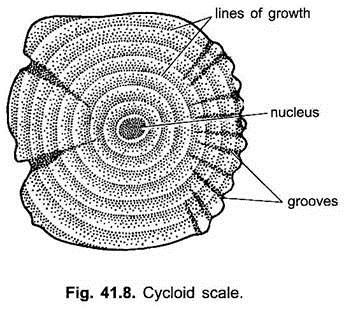
Cycloid scales (Fig. 41.8) are round, thick in the centre and thin towards the margins. They have a lower layer of fibrous connective tissue and an upper layer of bone-like isopedine which is elaborated to form dentine. They show concentric lines of growth which indicate the age of the fish. The scales lie embedded in the dermis diagonally overlapping each other.
The posterior part of each scale overlapping the anterior part of the scale behind, thus, covering the body with a double layer of scales. The exposed part (posterior) has a smooth edge, while the concealed part may have a wavy margin. Cycloid scales form a dermal exoskeleton in many bony fishes.
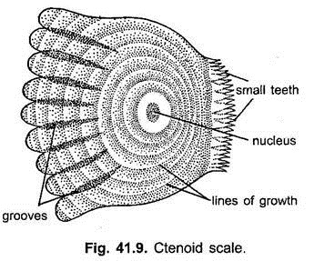
Ctenoid scales (Fig. 41.9) probably arose from the more simple cycloid scales. They have the same shape, structure and concentric lines as the cycloid scales, but they differ in having small teeth or cteni on their free posterior part. Their anterior concealed part may have notched or scalloped margin. Ctenoid scales form a dermal exoskeleton of most bony fishes.
The cycloid or ctenoid scales covering the lateral line canals are perforated by a vertical tube of the lateral line opening on the surface. In some flat fishes grooves both cycloid and ctenoid scales are present. In some catfishes there are no scales, in eels scales are very small and embedded in the dermis. In sea-horses scales form a continuous armour covering the body. Scales of bony fishes arise only from dermis, they form a protective exoskeleton but do not hamper movement.
In tetrapoda dermal scales are known as bony plates or osteoderms. In Apoda (Amphibia) only vestiges of osteoderms are present. They are found in pockets below the epidermis and are not visible externally. Two most ancient groups of reptiles, the turtles and crocodiles, have retained bony dermal plates.
Turtles have continuous osteoderms below the epidermal plates of the carapace and plastron. These osteoderms form a rigid dermal skeleton which becomes connected with the endoskeleton. In crocodiles there are osteoderms below the epidermal plates only on the back and the throat.
In birds and mammals there is a tendency for elaborating epidermal structures with an accompanied reduction or loss of dermal derivatives. In mammals, osteoderms are found in armadillos lying below the epidermal scales. They are bony plates of spongy texture. Extinct glyptodons had rigid bony armour of osteoderms. In some whales bony osteoderms may be present on the back and the dorsal fin.
