In this article we will discuss about the development of kidney in various vertebrates.
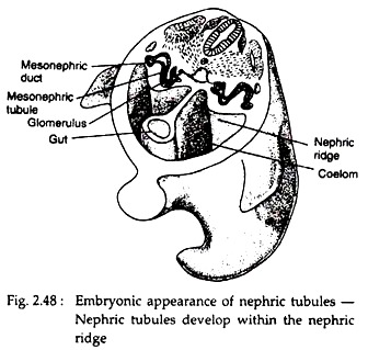
The kidneys develop within the postero-dorsal mesoderm of the embryo. At the onset of development this mesoderm expands to form a nephric ridge (Fig. 2.48). The next structure that appears is the paired nephrotome. The nephrotome is usually segmental and contains the nephrocoel, which is a coelomic chamber that may open via a ciliated peritoneal funnel into the coelom.
From the median part of nephrotome develops glomerulus and from the lateral end grows ducts. Several ducts fuses to form a common nephric duct. At this point the nephrotome is called nephric tubule, which is the fundamental structure of the urinary system.
The nephric tubule opens at one end into the coelom and on the other end into the nephric duct, with a glomerulus in between. In adult vertebrate kidney, the opening of nephric tubule in the coelome is rarely found. The fundamental organization is changed during development.
Tripartite Concept of Kidney Organisation:
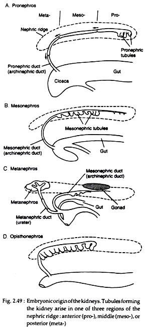
During development, the nephric tubules differ structurally within the nephric ridge. This difference is described by different authors as tripartite concept. According to this concept the nephric tubules develop in three regions of the nephric ridge.
Subsequent merger, loss or replacement of these tubules constitutes the developmental basis for the definitive adult kidneys. Nephric tubules that arise at the anterior region is called pronephros, tubules arise at the middle region is called mesonephros and which arise at the posterior region is called metanephros (Fig. 2.49).
(i) Pronephros:
ADVERTISEMENTS:
Tubule that develops within the anterior part of the nephric ridge is called pronephric tubules. Pronephros usually develops and retained for a short duration, during developmental stages of vertebrates. The pronephric tubules join to form a common pronephric duct (Fig. 2.49A).
This duct grows posteriorly and reaches and opens into the cloaca. The glomerulus protrudes from the roof of the coelom and filters fluid from coelom. Pronephric tubules then take up this coelomic fluid via the ciliated peritoneal funnels and excrete as urine.
Pronephric tubules and glomeruli form functional kidney in larval cyclostomes, in some adult fishes and in the embryos of most of the lower vertebrates. In most other vertebrates, the embryonic pronephros regresses, and as it does so, it is replaced by a second type of embryonic kidney, the mesonephros.
(ii) Mesonephros:
ADVERTISEMENTS:
Middle part of the nephric ridge gives rise to the tubules of mesonephric kidney (Fig. 2.49B). These mesonephric tubules do not form a separate new duct for excretion; instead tap into the existing pronephric duct. Now this duct is coined as mesonephric duct.
The mesonephros usually remains functional in the embryonic condition. But in some adult fishes and amphibians it persists. In that case, it is modified by the incorporation of additional tubules arising within the additional posterior tubules. This is termed as opisthonephros (Fig. 2.49D). In amniotes, the mesonephros is replaced in later development by a third type of embryonic kidney, the metanephros.
(iii) Metanephros:
In case of metanephros, metanephric duct appears first as metanephric duct, as a ureteric diverticulum, at the base of preexisting mesonephric duct. This ureteric diverticulum grows dorsally into the posterior part of the nephric ridge and stimulates the growth of metanephric tubules.
These tubules and ducts form the metanephric kidney. This type of kidney is present in the adult amniotes and the metanephric duct is called as ureter (Fig. 2.49C).
Kidney in Lower Vertebrates — Pro and Mesonephros:
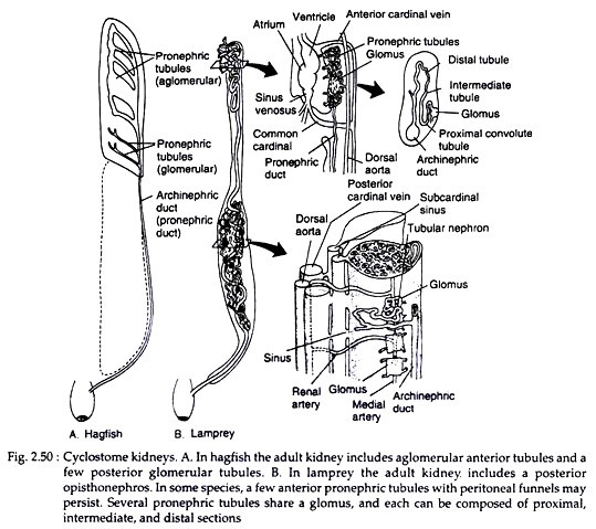
In hagfish, pronephric tubules unite successively with one another, forming a pronephric duct (Fig. 2.50A). Anterior tubules lack glomeruli but open to the coelome via peritoneal funnels. Posterior tubules are associated with the glomeruli but lack connection to the coelom.
In adult, aglomerular tubules together with several glomerular tubules become the compact pronephros. This pronephros contribute to the formation of coelomic fluid. However, in adult hagfish mesonephros is the functional kidney.
In lampreys, the ammocoetes larva possesses pronephric kidney, consisting of three to eight coiled tubules served by a single compacted bundle of capillaries called a glomus. A glomus differs from a glomerulus in that each vascular glomus services several tubules (Fig. 2.50B).
Kidney in Amphibians — Opisthonephros:
In amphibians, early embryonic pronephros is usually succeeded by the larval mesonephros, which upon metamorphosis is replaced by an opisthonephros. The anterior kidney tubules also transport sperm in adult, illustrating the dual use of the ducts that serve as both genital and urinary system.
Kidney in Other Vertebrates:
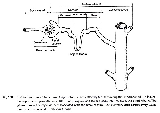
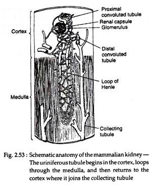
In most of the amniotes, the mesonepnros is completely replaced by metanephros in adults. The metanephros drain its products by a new urinary duct, the ureter. A metanephric tubule is long and well differentiated into proximal, intermediate and distal regions. In mammals, in particular, the intermediate section of the tubule is specially elongated, constituting the major part of the loop of Henle (Fig. 2.52 and 2.53).
Loop of Henle:
The loop of Henle of the metanephric tubule occurs only in the group of animals who are capable of producing concentrated urine. Among vertebrates only mammals and birds can produce urine, which is much concentrated than their blood. Therefore, loop of Henle is present in these groups only. The ability to produce concentrated urine is directly correlated with the length of the loop.
The term loop of Henle refers to both a positional and structural feature of nephron. Positionally, the loop refers to the part of the tubule which comes out of the cortex region and enters into the medulla of the kidney. The entering part of the tubule, from glomerulus, to medulla, is called descending limb. The outgoing part from the medulla to cortex is called the ascending limb (Fig. 2.53).
Structurally, the loop of Henle has three regions — these are:
(i) The straight portion of the proximal tubule.
(ii) The thin-walled intermediate region.
(iii) The straight portion of the distal tubule. Except the intermediate region, the other two regions are thick-walled. The terms thick and thin refer here to the height of the epithelial cells forming the loop. Cuboidal cells are thick and squamous cells are thin.
