In this article we will discuss about the circulatory system in vertebrates with the help of suitable diagrams.
The circulatory system in craniates differs basically from that of the invertebrates. A progressive evolution in this group has led to a gradual change, leading to the appearance of an efficient heart, since activities of animals are directly related to a rapid flow of oxygenated blood.
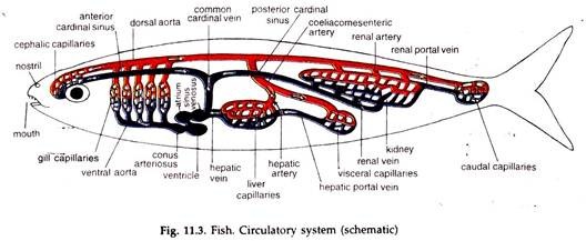
The general scheme of circulation, sending blood through arteries and returning it to the heart through veins, has been complicated by the interposition of gills in the course of outflow of blood from the heart, and the arteries are to function as both afferent (sending away) and efferent (returning) vessels in branchiate craniate (Fig. 11.3).
Flow of oxygenated blood to the tissues directly from the gills and returning deoxygenated Mood from tissues to the heart created the necessity of imposition of some blood purifying organ in its course.
ADVERTISEMENTS:
In the course of returning current, the liver and the kidneys have been imposed and portal systems developed. Both renal and hepatic portal systems continue to present in amphibians and reptiles; only the hepatic portal system is retained in birds and mammals.
From a single atrium, single ventricle heart in branchiate chordates to two atria, one ventricle heart in amphibians and reptiles, and two atria, two ventricle heart in birds and mammals have evolved in response to the need of a more and more efficient heart.
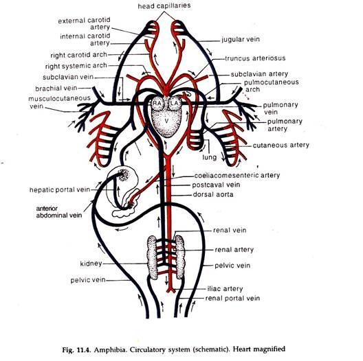
Replacement of gills by lungs in terrestrial vertebrates has caused big changes in the heart and aortic arches. Blood started moving through different routes—deoxygenated blood from heart to lungs for oxidation and back to heart, and oxygenated blood from heart to tissues (where it loses oxygen) and back to heart, the latter being (Fig. 11.4) intervened by both the liver and the kidneys or by the liver alone.
Absence of any partition in the ventricle in three-chambered heart of amphibian permits mixing of deoxygenated and oxygenated blood and this stands in the way of leading an active life in this group.
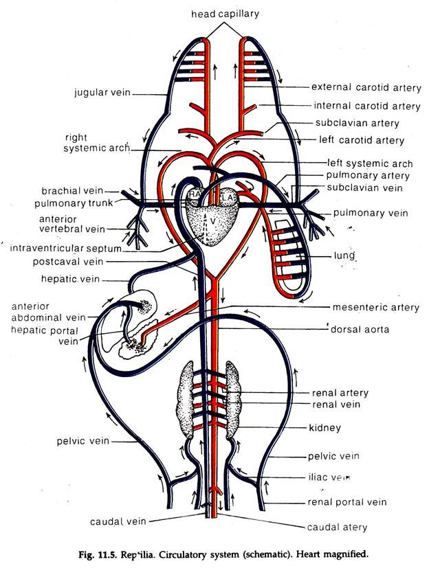
In reptiles, an incomplete partition in the ventricle (Fig. 11.5) restricts mixing of two bloods to a certain extent. In crocodiles, a group of reptiles, the partition is near complete and the mixing of blood is prevented to a great extent, enabling them to lead more active life.
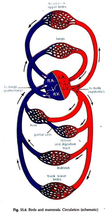
In birds and mammals, a four-chambered heart (Fig. 11.6) has ensured complete separation of oxygenated and deoxygenated blood and this has helped them to lead very active life.
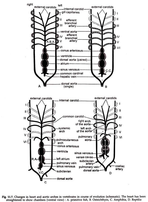
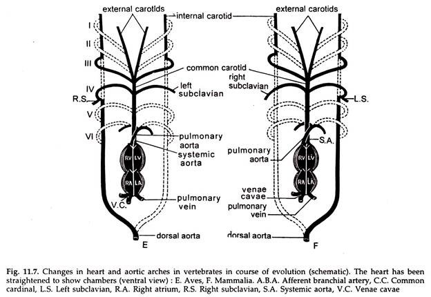
With the progress of evolution a gradual reduction in the number of aortic arches occurred in craniata. From eight in cyclostomes it came down to six in primitive fishes, to four in Osteichthyes. With the adaptation to land life the number of arches drastically reduced to three in tetrapod’s (Fig. 11.7). Their embryos, however, always possess six arches.
Twenty per cent of the atmospheric air being oxygen, the land forms had to move a far less amount of air to respiratory membranes, than the amount of water to be moved in aquatic forms. In primitive tetrapod’s (frogs and toads) the sixth pair formed the pulmonary arteries, their bases constituting the pulmonary arch, took over the function of carrying deoxygenated blood from the heart to lungs and skin.
This is maintained (skin excluded) in higher groups of vertebrates. The anterior two pairs degenerated greatly, the rudiments of their ventral portions formed a part of the ventral branches of the external carotids. The anterior portion of the ventral aortae became external carotids. The third pair formed the carotid arches. The anterior portion of the dosal aortae formed internal carotids.
The part of the ventral aortae between the bases of the third and fourth arches became common carotids. The fourth pair formed systemic arches; the fifth pair degenerated. Amphibians and reptiles retained paired systemic arches.
Asymmetry in the systemic arches occurred in birds and mammals. The left systemic arch degenerates in birds, while the right one degenerates in mammals. In mammals the right subclavian artery arises from the right systemic arch before it is reduced during development.
The change from somewhat sluggish life in most invertebrates to active life of vertebrates created a demand for separation of blood from coelomic fluid and also a rapid circulation. In response to this, the open circulatory system changed to a closed circulatory System.
ADVERTISEMENTS:
The closed circulatory systems are of two types:
1. Single circulation:
To complete one cycle, i.e. to return to the starting point, blood flows only once through the heart. This is found in branchiate craniates. Heart gills → tissues heart.
2. Double circulation:
To complete one cycle blood travels twice through the heart. This is present in terrestrial chordates. Heart (left ventricle) tissues heart (right ventricle) lungs → heart (left ventricle).
Heart function:
Irrespective of the number of chambers, the primary function of the heart is to send blood in an orderly manner to the tissues and to receive the blood back. This is met with in vast majority of animals by having a muscular heart and vessels. The blood is received by the atrium/atria and pumped out by the ventricle(s).
Valves are present at the openings in the heart and the opening of the aorta to prevent backflow of blood and thereby ensure a steady, one-way movement. In addition the thick walls of the arteries can contract and the veins, being thin walled, are provided with valves at regular intervals.
The pressure exerted by the ventricle is sufficient to drive blood through arteries and its return to the heart. In some arthropods the blood pressure falls below the thrust required to drive the haemolymph into the heart. The pericardial sinus enclosed in the pericardium is filled with haemolymph.
Contraction of muscle stands extending from the heart to the pericardium causes expansion of the heart and the haemolymph in pericardial sinus is sucked into the heart through Ostia in the heart wall. The fall of pressure of fluid in the pericardial sinus draws in haemolymph from the large sinuses. Such a heart is known as suction heart.
Normally, both blood pressure and the blood flow rate are lowest in the capillaries. Still they together are sufficient to drive the blood through veins. An increase in blood flow rate, once it enters the venules help in the process. Valves in the veins prevent back-flow of blood.
The ventricle and the atria contract and relax in successions. A heart beat consists of two steps, the contraction or cystole and relaxation or diastole. The total number of heart beat per minute is heart rate. This corresponds to the pulse. The ventricles contract with a great force driving the blood to the arteries.
The ventricles relax, pressure within it falls and blood from the atria enters it. The atria contract when the ventricles are near full with blood. This is again followed by the contraction of the ventricles. The alternate stretching and contraction of arteries are associated with the contraction and relaxation of the ventricles. This can be felt as pulse.
Cardiac Output:
Outflow of blood or haemolymph from the heart in per unit time is called cardiac output. Small animals, the animals under exercise need increased cardiac output.
Since the weight of heart in most mammals is about 0.6 per cent of the body weight, the cardiac output is increased by increasing heart rate. But the increased rate is not proportional as the blood carries more oxygen at such time. The pulse rate of an elephant is 25 beats per minute, of an adult man 70 and that of a shrew 600.
