In this article we will discuss about the digestive system of vertebrates with the help of suitable diagrams.
Embryonic Digestive Tract:
Archenteron:
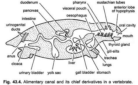
The embryonic archenteron becomes the lining of the adult digestive tract and of all its derivatives. Splanchnic mesoderm adds layers of connective tissue and smooth muscles around the archenteron. Ectodermal invagination of the head forms the stomodaeum leading into oral cavity, and a similar mid-ventral ectodermal invagination forms proctodaeum, which leads into the hindgut.
The stomodaeum becomes the adult buccal cavity and gives rise to teeth enamel, epithelial covering of tongue, glands, e.g., mucous, poison and salivary, etc., and Rathke’s pouch of anterior pituitary gland. The proctodaeum forms either a small terminal part of the cloaca in lower vertebrates and rectum in mammals.
The alimentary canal in embryos from stomach to cloaca is attached to the dorsal body wall by a double fold of peritoneum, called the dorsal mesentery, and to ventral body wall by a ventral mesentery. In adults, dorsal mesentery persists but the ventral mesentery disappears leaving only in the region of liver and urinary bladder.
Digestive Tract of Adult:
The digestive tract differentiates for different functions into the following regions- mouth, buccal cavity, pharynx, oesophagus, stomach, small intestine, large intestine and cloaca. Following outgrowths arise from the digestive tract- oral glands, Rathke’s pouch, thyroid gland, gill-clefts, tympanic cavity, thymus and other glands of gill-clefts, trachea, lungs, swim bladder, liver, pancreas, yolk sac, and urinary bladder.
Histology:
The wall of the alimentary canal is made of four concentric layers.
ADVERTISEMENTS:
They are:
(i) An outermost visceral peritoneum or serous coat is made of mesothelial cells and thin layer of connective tissue. It is lacking in the oesophagus,
(ii) Below this is a muscular layer formed of smooth muscle fibres arranged in outer longitudinal and inner circular muscle fibres. Between the two layers of muscles is a network of nerve cells and nerve fibres of the autonomic nervous system, known as myenteric plexus or plexus of Auerbach.
(iii) Beneath the muscle layer is a submucosa made of connective tissue having elastic fibres, fat, blood and lymph vessels, nerve cells and fibres glands,
ADVERTISEMENTS:
(iv) The innermost layer is a mucosa composed of three regions:
(a) Outer-most narrow muscularis mucosa of outer longitudinal and inner circular smooth muscle fibres.
(b) Middle thin layer of lamina propria of connective tissue, blood vessels, nerves and nodules of lymphatic tissue, and
(c) A basement membrane supporting a layer of columnar epithelial cells which are often glandular and ciliated.
Mouth:
Mouth is the opening leading into buccal cavity. In lampreys (cyclostomes) it is a circular opening at the base of buccal funnel and remains permanently open due to lack of jaws, etc. In gnathostomes it is terminal. Mouth is bounded by lips which are immovable and formed of cornified skin in fishes, amphibians and reptiles. In mammals these are fleshy and muscular.
Buccal Cavity:
The space between the lips and the jaws is a vestibule. It may be bounded on the outside by cheeks and on the inside by the gums. Mucous glands of cheeks open into the vestibule. The mouth opens into a buccal cavity, which is a space between the mouth and the pharynx. The exact point where the stomodaeal ectoderm and pharyngeal endoderm merge is variable and not easy to discern.
ADVERTISEMENTS:
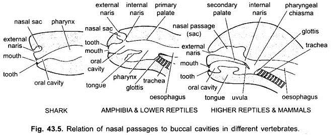
In elasmobranchs and most bony fishes the nasal cavities do not open into the buccal cavity. In Chondrichthyes and tetrapoda (amphibians and most reptiles) the nasal cavities open into the buccal cavity by choanae or internal nares, which are primitively placed anteriorly, but in crocodiles, birds and mammals they become posterior in the pharynx due to the formation of a secondary palate, which effectively separates the respiratory nasal passage from the mouth cavity or food passage.
In birds, this palate is cleft due to which nasal and buccal cavities communicate with each other. In mammals, secondary palate is continued posteriorly as a membranous soft palate. In human beings soft palate hangs into the laryngeal pharynx in the form of fleshy process, called uvula.
Derivatives of Buccal Cavity:
1. Oral Glands:
There are two kinds of integumetary multicellular glands opening into the buccal cavity. They are mucous glands and enzymatic glands. Fishes and aquatic amphibians have only mucous glands. Reptiles have glands in groups, such as palatine, lingual, sublingual, and labial glands named according to location, they also produce mucus.
In poisonous snakes the upper labial glands are modified to secrete venom, while in the Gila monster the sublingual glands produce poison. Birds have sublingual glands and a gland in the angle of the mouth. Mammals have many small mucous glands besides true and enlarged salivary glands which are enzymatic. They are parotid, sublingual, submaxillary and infraorbital salivary glands, secreting mucin and ptyalin.
2. Tongue:
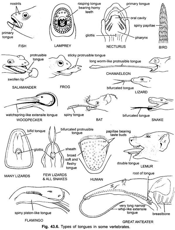
The tongue is found mostly in all vertebrates. Tongue in vertebrates show much diversity and are not homologous. In cyclostomes, there is a muscular, fleshy, rasping tongue with horny teeth for rasping the skin and muscles of their prey.
Fishes have a primary tongue formed of a fleshy fold of the buccal floor. It has no muscles, but receptors and teeth are present on the tongue in some bony fishes. The tongue is covered with mucous membrane. In some amphibians the tongue is either lacking or immovable. Most amphibians, however, have a protrusible tongue and in some frogs and toads it may be folded back on itself when not in use.
It can be thrown out of mouth by rapid inflow of lymph for capturing the insects. The tongue in lizards and snakes is often highly developed. In chameleons, it is very extensible used to capture insects. The tip is thickened and sticky. The forked tip of the tongue in snakes serves as a means of transferring chemical stimuli from the external environment to the paired vomero-nasal organs on the roof of the mouth.
In turtles and crocodilians, the tongue cannot be extended. The amniote tongue has voluntary muscles, it receives the hypoglossal nerve and has glands and taste buds. It also develops intrinsic muscles which move the tongue. In birds, the tongue is slender and has a horny covering. In some birds the tongue is immobile, while in some birds it is long, protractile and often used for capturing the food.
In most mammals, except whales, the tongue is highly developed and capable of considerable movement, in addition to extension and retraction, due to the presence of a number of intrinsic muscles.
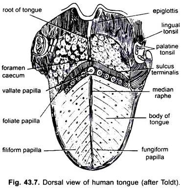
In mammals the mucous membrane below the tongue forms a median fold, called frenulum which joins the tongue to the floor of the mouth. In mammals the upper surface of the tongue bears four kinds of papillae, (filiform, fungiform, foliate and circumvallate), bearing taste buds except filiform papillae.
3. Teeth:
Vertebrates have two types of teeth attached to jaw bones- epidermal teeth and true teeth. Epidermal teeth are best developed in cyclostomes. They are hard, conical, horny structures derived from the stratum corneum. In lampreys they are found on the walls of the buccal funnel and on the tongue. Tadpole larvae of frogs and toads have serrated epidermal teeth in rows on the lips. In mammals the adult duckbill platypus has epidermal teeth.
True Teeth:
Teeth are not found in baleen whales and anteaters in mammals, and agnathans, sturgeons, some toads, sirenians, turtles and modern birds, etc. In lower vertebrates (such as fish, amphibians and most reptiles) teeth may be replaced continually an indefinite number of times, such teeth are called polyphyodont. These teeth are homodont (similar type) and acrodont. (with the jaw bones).
In most mammals teeth are diphyodont, thecodont and heterodont. In some mammals these are monophyodont having only one set of teeth, e.g., moles, Indian squirrel. Teeth are similar in structure to the placoid scales of sharks, formed of a central pulp cavity, around which is present a thick but soft layer, the dentine, which is externally covered by a thin, extremely hard enamel. These are supposed to have derived from bony scales of ostracoderms and placoderms. For details readers may see the Dentition in Mammals.
4. Adenohypophysis:
The anterior lobe of pituitary gland develops as a dorsal evagination of stomodaeum, called Rathke’s pouch, which constricts off to form the anterior and middle lobes of pituitary gland (adenohypophysis). The posterior lobe of pituitary or neurohypophysis is the ventral evagination of diencephalon, called infundibulum. Thus, it is nervous part.
Pharynx:
The part of the alimentary canal immediately behind the buccal cavity is a pharynx, lined with endoderm. It is a common passage serving both for the digestion and respiration. As a part of the digestive system it is used as a passage for food from the buccal cavity to the oesophagus, its muscles initiate swallowing.
In fishes, the pharynx is large and laterally perforated for gill-slits, while in tetrapoda, it is short and bears openings of nostrils. In embryos, the wall of pharynx gives off a number evaginations which develop into spiracles, gill-clefts, air bladders, lungs, tonsils and a few endocrine glands (e.g., thymus, thyroid and parathyroids).
Oesophagus:
The oesophagus is short in most fishes and amphibians because they lack neck, but it is longer in amniotes due to presence of neck. The oesophagus of reptiles is more elongate than that of fishes and amphibians. In granivorous and carnivorous birds, a portion of the oesophagus is enlarged into a sac-like pouch called crop which serves to store food which has been eaten quickly.
The crop is essentially lacking in digestive glands although in pigeons the crop has 2 crop glands in both sexes, they are really not glands but cell-forming structures, the cells form ‘pigeon milk’ which is fed to the young. In mammals, the oesophagus is long, lacks glands and varies in relation to the length of neck.
It passes through the diaphragm, the portion below the diaphragm is covered with visceral peritoneum which is lacking from the upper part. Oesophagus has mucous glands. Its lining forms longitudinal folds, or finger-like fleshy papillae (elasmobranchs) or horny papillae in marine turtles.
Histologically, the oesophagus differs from the rest of the alimentary canal in three facts:
(i) It has no visceral peritoneum because it lies outside the coelom, its outermost covering layer is a thin tunica adventitia.
(ii) The muscle fibres in its anterior part are striped, middle part has both striped and unstriped muscles, and the posterior part has only unstriped muscles. But there are exceptions in ruminating mammals, all the muscles are striped or voluntary.
(iii) The mucous membrane lining is made of stratified squamous epithelial cells and not of columnar cells.
Stomach:
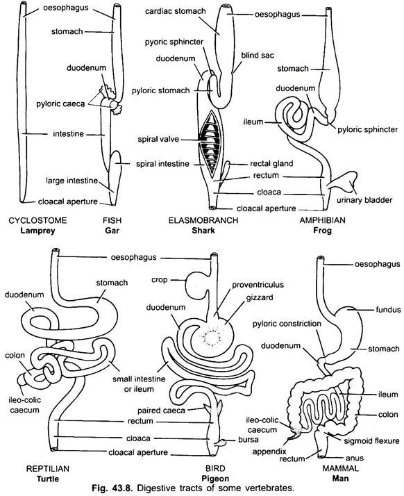
There is practically no stomach in cyclostomes, chimeras, lung fishes and some primitive teleost fishes, since it has no gastric glands, but in most fishes and tetrapoda it is dilated for storage and maceration of solid food, and digestion of food because it contains gastric glands.
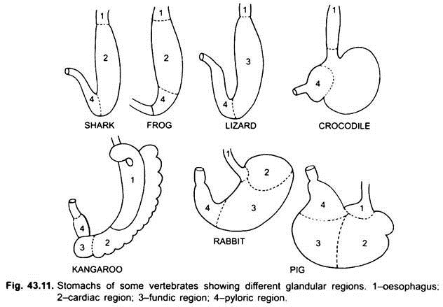
The first part of the stomach, next to the oesophagus, is the cardiac region and the lower end near the intestine is the pyloric region, which has a pylorus or pyloric valve in which the mucous membrane lining is surrounded by a thick sphincter muscle which regulates the opening and closing of the pyloric stomach into the intestine.
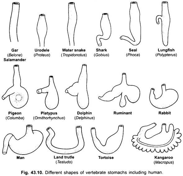
Stomach is straight in cyclostomes, gar, Belone, etc., and spindle-shaped in Proteus, Necturus, some lizards and snakes. In turtles and tortoises, it is a wide curved tube, and in elasmobranchs the stomach is J-shaped. In crocodiles and birds the stomach has two parts, a proventriculus with gastric glands, and a highly muscular gizzard, which represents the pyloric region and has a hard, cornified lining for grinding food.
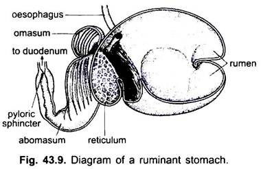
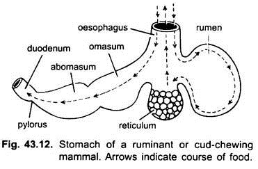
In mammals the stomach lies transversely and may be a simple sac or divided into 3 regions, namely cardiac, fundic and pyloric and each region has its gastric glands. In many ruminants the stomach has four chambers- a rumen, reticulum, omasum and an abomasum. It is claimed that the first three chambers are modifications of the oesophagus, and abomasum is the true stomach representing the cardiac, fundic, and pyloric parts of the stomach.
It has been shown embryologically that all four chambers are modified regions of the stomach. In camels, there is no omasum, the rumen and reticulum have pouch-like water cells which were once believed to store water, but they are probably digestive.
Histologically, the stomach has the typical parts of the alimentary canal, but it has two peculiarities, the muscularis mucosa is made of an outer longitudinal layer and an inner circular layer of muscles. The epithelium lining is thick with several types of glandular cells forming gastric glands of three types called cardiac, fundic, and pyloric gastric glands.
The cardiac and pyloric glands secrete only mucus from their surface cells. Fundic glands (or cardiac glands in some) have three kinds of cells, mucous neck cells produce mucus, oxyntic cells produce hydrochloric acid, they may be present in the cardiac region also, zymogen cells or peptic cells secrete pepsin.
In most animals the zymogen cells also secrete two proenzymes called propepsin and prorennin which are converted by hydrochloric acid into pepsin and rennin respectively. The secretions of all stomach cells form a mixture called gastric juice.
Small Intestine:
Small intestine is long, narrow and coiled tube after the pylorus. It is the most important part of the digestive tract because the digestion and absorption of food take place in it. In cyclostomes, the intestine is a short straight tube with a spirally arranged longitudinal flap extending into it.
In elasmobranchs, it is divided into small and large portions, and the small portion has a spiral valve which greatly increases the absorptive surface. A spiral valve is also present in the small intestine of a few more primitive bony fishes, but is lacking in higher forms in which the intestine is long and coiled.
In caecilians, it is little coiled and not differentiated into a small and large tract. In frogs and toads it is relatively long and coiled. In reptiles, it is more coiled than in amphibians. For the first time in vertebrates a caecum or blind diverticulum arises at the junction of small and large intestines.
However, this is not permanent in all reptiles. In birds, the small intestine is coiled or looped and one or two colic caeca are also present at the junction of small and large intestines. In most mammals also the small intestine is proportionately long and coiled. Its length, however, is correlated with food habits. In herbivores it is relatively more longer in comparison to insectivores and carnivores.
There is a blind pocket or caecum at the junction of the colon and small intestine which is generally small in carnivorous species and quite long in many herbivores. The first part of the small intestine is the duodenum, which is short beginning from pylorus and terminates beyond the entrance of pancreatic and hepatic ducts.
It has many folded villi and contains branching Brunner’s glands in the submucosa which secrete mucus, some alkaline watery-fluid, and a little enzyme. Duodenum also produces two hormones called secretin and cholecystokinin which stimulate the pancreas and gall bladder to liberate their juices. Ducts from the gall bladder and pancreas open into the duodenum.
Behind the duodenum is an ileum, which only in mammals is differentiated into an anterior smaller jejunum and posterior longer ileum. A large number of small digestive glands are present in the small intestine. They are tubular glands or crypts of Lieberkuhn found through the entire length, they secrete mucus and a succus entericus which has several enzymes.
The lining of the small intestine is folded to form small villi, which increase the surface area for secretion and absorption. The villi are covered densely by minute finger-like projections, called microvilli which assist in absorption into the villi. In mammals nodules of lymphoid tissue called Peyer’s patches are found on the ileum.
Large Intestine:
Large intestine has a larger diameter than the small intestine. It is generally short in fishes, amphibians, reptiles, and birds, but in mammals it is long. In lower forms the large intestine forms a rectum, but in tetrapoda it has a colon and terminal rectum. In most fishes and amphibians, the terminal part of the rectum leads into a cloaca formed by the proctodaeum.
The rectum, excretory ducts, and genital ducts open into the cloaca, and it opens to the exterior by a cloacal aperture. But in many bony fishes and all mammals (except monotremes) the rectum and urinogenital ducts have separate openings to the exterior; the opening of the former is an anus.
Rectum of mammals is not homologous with the rectum of vertebrates since in mammals it is derived by partitioning of embryonic cloaca. In most vertebrate embryos there is a postanal gut as an extension of the intestine into the tail, but it disappears later.
In elasmobranchs the large intestine bears a pair of rectal glands which secrete mucus and sodium chloride. In amniotes there is an ileocolic valve at the junction of small and large intestines, which is absent in fishes. It prevents bacteria to enter into ileum from colon.
In amniotes from this junction arises an ileocolic caecum which is two in birds. It contains cellulose digesting bacteria. It is very long in herbivorous mammals (rabbit, horse, cow, etc.). In primates caecum is small having a vestigial vermiform appendix.
Digestive Glands:
1. Liver:
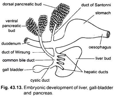
The liver arises as a single or double outgrowth from the ventral wall of the embryonic archenteron. This outgrowth forms a hollow hepatic diverticulum, which soon differentiates into an anterior part, which proliferates to become the liver and its bile ducts, and a posterior part, which gives rise to the gall bladder and cystic duct. The bile ducts join to form a hepatic duct which unites with the cystic duct to form a common bile duct or ductus choledochus. The region of the archenteron from which the liver arises becomes the duodenum.
The liver is the largest lobed gland in the body, suspended by a double layer of peritoneum from the transverse septum or its representative.
A gall bladder is for storage of bile secreted by the hepatic cells, lies in the liver and drains into the duodenum through common bile duct formed by the union of cystic duct and hepatic duct. A gall bladder is not indispensable and is lacking in many birds and mammals.
A liver is present in all vertebrates. In cyclostomes, it is small, single lobed (lampreys) and two lobed in hagfishes. It is bilobed in elasmobranchs, two or three lobed in bony fishes, amphibians, reptiles and birds and many lobed in mammals. Liver is long, narrow and cylindrical in fishes, urodeles and snakes.
It is short, broad and flattened in birds and mammals. A gall bladder and bile duct are present in larval cyclostomes but they are absent in the adult. Fishes, amphibians, and reptiles generally have a gall bladder, but it is lacking in many birds. Most mammals have a gall bladder, but it is absent in Cetacea and Ungulata.
The liver secretes a watery, alkaline bile but have no enzymes. It neutralises the acidity of food entering the duodenum. It aids in the digestion of fats.
2. Pancreas:
Pancreas is formed from the endoderm of the embryonic archenteron. A single dorsal diverticulum from embryonic duodenum and one or two ventral outgrowths from the liver form pancreatic diverticula. The proximal parts of diverticula form pancreatic ducts, but these ducts undergo reduction or fusion so that only one or two pancreatic ducts remain in the adult, they open into the duodenum either separately or after uniting with the common bile duct.
The distal parts of diverticula undergo budding to form the main mass of pancreatic cells to which mesodermal derivatives are added. Thus, a single gland is made which has several lobes forming either a diffuse or a compact pancreas.
The pancreas is both an exocrine and endocrine gland, bound together by delicate strands of connective tissue. The exocrine part secretes digestive enzymes which are poured into the duodenum through pancreatic ducts. Whereas the endocrine part secretes hormones such as insulin and glucagon.
A pancreas is present in all vertebrates. In lampreys, some bony fishes, lungfishes and lower tetrapods, it is a diffuse organ embedded in the liver, mesenteries and intestinal wall. Hagfishes have a small pancreas. Elasmobranchs have a well defined bilobed pancreas. In higher tetrapoda it is generally a compact gland. One or two pancreatic ducts open into the duodenum.
