Gonads are cytogenic sex glands. They are paired testes in males and ovaries in females because all vertebrates are dioecious with the exception of the hermaphrodite cyclostomes and some bony fishes. In hagfishes the anterior part of gonad produces ova and the posterior part sperm.
In any one individual, however, the gonad at maturity functions only as an ovary or testis, not as both. The gonads are not paired in cyclostomes and ducts are absent. The ova or sperm pass directly into the coelom and to the outside through genital pores.
Most bony fishes that are functionally hermaphroditic are marine, although a few are freshwater, such as killifish (Rivulus marmoratus) and the Asiatic synbranchid (Monopterus albus). Some are like the hagfish, they function first as one sex, and then transform into the opposite sex. Other possess ovotestes in which the mature ova are fertilised by sperm produced within the same organ before they are laid.
The mesodermal coelomic epithelium forms a pair of thick, elevated, elongated genital ridges lying longitudinally along the dorsal surface of the coelom near the developing kidneys. Genital ridges are much longer than the adult gonads. The middle part of mesoderm of genital ridges gives rise to the gonads, whereas the anterior (progonal) and posterior (epigonal) parts remain sterile.
ADVERTISEMENTS:
While the peritoneum covering them becomes the germinal epithelium. The gonads, thus, formed become suspended from the dorsal body wall by bands of tissue known as mesorchium in the male and mesovarium in the female.
The germinal epithelium of ovaries lies on their outer surface and gives rise to ova, consequently the ova are discharged from the surface into the coelom. The germinal epithelium of testes lines their seminiferous tubules, hence, spermatozoa are released into the lumen of seminiferous tubules.
Testes and Male Genital Ducts:
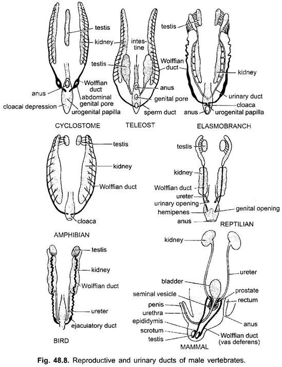
Cartilaginous and bony fishes have paired gonads and the sexes are generally distinct. In cyclostomes gonads are unpaired and ducts are absent. Testes are paired in other vertebrates and are usually found attached to the kidneys. A testis is a compact gland covered over by coelomic epithelium and formed of a mass of highly coiled seminiferous tubules in connective tissue.
These are lined by the germinal epithelium. In dogfish, the testes are elongated structures. In anamniotes the anterior portion of the mesonephric kidney comes to serve the male genital system and the posterior part as renal organ. This anterior portion loses its peritoneal funnels and Malpighian bodies and its uriniferous tubules loose excretory function and form vasa efferentia which become continuous with seminiferous tubules of adjacent testis.
ADVERTISEMENTS:
Thus, the mesonephric kidney of adult anamniotes has an anterior genital portion and a posterior renal portion. The mesonephric duct is a urinogenital duct in male, it serves both as a urinary duct for urine and as a vas deferens for sperms.
But in many elasmobranchs (e.g., dogfish) special accessory urinary ducts are formed, one for each kidney to drain urine from kidney to cloaca and the mesonephric duct is only genital in function and is a vas deferens. The anterior genital part of kidney with a part of mesonephric duct forms the epididymis.
In amphibians, testes are paired and are connected either directly or by way of mesonephric tubules to the archinephric duct which in turn opens into the cloaca. No special copulatory organs are present. In some toads there is a structure called Bidders organ located anterior to each testis. Under certain circumstances this may develop into an ovary.
ADVERTISEMENTS:
In male amniotes the adult functional kidneys are metanephroi, each has its own ureter for conducting urine, hence, the persistent mesonephric duct is taken by the testis and becomes an entirely genital vas deferens or deferent duct. Its upper end is greatly coiled and forms a compact structure called the epididymis. In many species of reptiles, special copulatory organs for the transference of sperm to the females are present.
In lizards and snakes, a pair of extrusible structures in the cloaca called hemipenes is present, while crocodiles and turtles possess a structure that may be homologous with the mammalian penis. In ducks and geese, a single penis-like structure, similar to that of turtles is present.
It is derived from the antero-ventral wall of the cloaca. The mammalian testes are either situated posteriorly in the body or they are outside the body cavity in a sac called the scrotum. In certain species testes descend into the scrotal sac only during the breeding season. The male possesses a single penis, which in monotremes is located on the floor of the cloaca.
Ovaries and Female Genital Ducts:
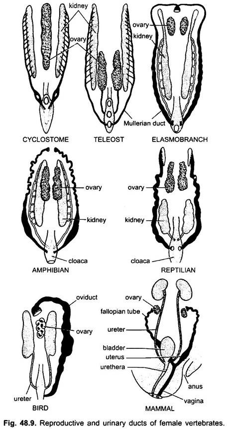
The ovaries are generally masses of connective tissue within an outer germinal epithelium. In the ovary are ova in various stages of development formed from germinal epithelium. The ovaries are never connected with the kidneys, nor are they connected with their ducts in most vertebrates, hence, ova pass into the coelom after being discharged.
In female anamniotes a coelomic funnel is formed from the nephrotome, the funnel grows back to form a groove on each side. The groove becomes closed to form a tube known as oviduct or Mullerian duct which lies on the outer side of the mesonephric duct and runs backwards to join the cloaca.
A Mullerian duct also appears in the male, but it is suppressed by androgens and it soon becomes vestigial. Thus, there are two ducts on each side in female anamniotes, a Mullerian duct acting as an oviduct and a mesonephric duct serving as a ureter.
But in most elasmobranchs the original pronephric duct splits longitudinally to form two ducts, one becoming the mesonephric duct for urine and the other forming an oviduct for ova which appropriates one or more peritoneal funnels to form its opening into the coelom. The method of formation of elasmobranch oviduct is considered to indicate its origin in vertebrates.
In cartilaginous and bony fishes, the female usually has two oviducts. In elasmobranchs the upper ends of these oviducts are fused so that there is a single funnel-like opening or ostium into which eggs from the ovaries may pass. Also in this group each oviduct possesses a shell gland, since the eggs are fertilised internally and then encased in a horny shell.
In most vertebrates the ovary is not directly connected with the oviduct so that the eggs enter the coelom and then pass into the ostium. In some bony fishes, however, where huge number of eggs is produced during the short breeding season, the ovaries are continuous with the oviducts so as to prevent the ova from escaping into the coelom. In many teleosts the lower parts of the paired oviducts are fused. Most bony fishes are oviparous, but there are some in which eggs hatch internally.
The amphibian ovaries are paired and contain a cavity within them that is filled with lymph. The oviducts are also paired although in some forms the lower ends are fused together. Frequently the lower end of each oviduct is enlarged into a uterus-like structure or ovisac which serves as a temporary storage space for ova before they are shed. Glands that secrete a jelly-like covering for eggs are usually situated along the oviduct.
In reptiles also paired ovaries contain cavities filled with lymph as in amphibians, or may be solid as in birds and mammals. Paired oviducts open separately into the cloaca. The albumen and shell producing glands are located along the oviducts. Archinephric or Wolffian duct degenerates in female reptiles.
In most birds the right ovary and oviduct, although present in embryonic development, become vestigial so that only the left genital system is functional. Along the course of oviduct are several glands which secrete membranes over the eggs, including layers of albumen, shell membranes and a calcareous shell.
In female amniotes the development of mesonephric and Mullerian ducts is the same as in anamniotes, but the adult kidneys are metanephroi, each with its own ureter, hence, mesonephros and its duct degenerate leaving only vestiges. The Mullerian duct acts as an oviduct and the metanephric duct as the ureter on each side.
In most vertebrates an oviduct is not differentiated into parts, and the two oviducts remain separate and open into the cloaca, but in mammals each oviduct is differentiated into two or three parts- (i) a narrow anterior Fallopian tube, (ii) wide posterior uterus whose posterior part may be differentiated as a vagina. In monotremes the two oviducts are separate so that there are two separate uteri opening into the cloaca. Monotremes have no vagina. With the separation of urinary and genital systems from the rectum, the cloaca is eliminated in marsupials and eutherians.
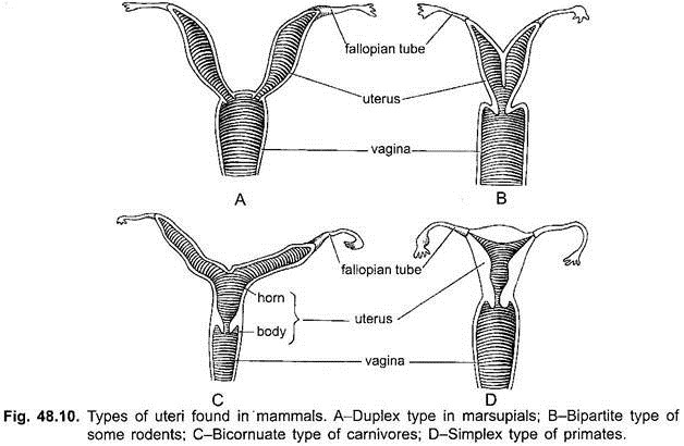
In marsupials there are two uteri (hence, Didelphia), and the terminal portions of oviducts are differentiated as vaginae. The two vaginae are separate but joined along the median line. They receive the penis of the male. But in eutherian mammals the two vaginae fuse into a single tube.
The two uteri may fuse partly or completely, thus, four kinds of uteri are recognised in Eutheria:
1. Duplex uterus found in rodents, elephants, and some bats. The two uteri do not fuse but enter and open separately in the vagina.
2. Bipartite uterus found in carnivores, pigs and cattle. The lower ends of the two uteri fused together so that they open by a single aperture (os uterus) into the vagina.
3. Bicornuate uterus found in ungulates, whales and most bats. The lower ends of the two uteri fused more completely with no partition wall between the fused portions and this part has a single cavity.
4. Simplex uterus found in Primates. The two uteri fused completely forming a single body with one cavity. There is no correlation between the development of various types of uteri and the phylogenetic relationship between the orders of mammals.


