In this article we will discuss about modification of aortic arches in various vertebrates.
Modification in Fishes:
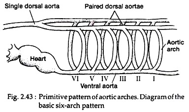
Branches from ventral aorta produce aortic arches, which divide into capillary beds within the gills. The part of the aortic arches delivering blood to the gills is called afferent branchial artery. From the dorsal end of the capillary bed arises another artery, called efferent branchial artery, which joins the dorsal aorta.
The capillary beds partially or completely encircle the gills and empty first into the collecting loop that joins the efferent artery (Fig. 2.43).
ADVERTISEMENTS:
The first afferent artery, which is expected to supply first pharyngeal slit, goes to a vascular sprout adjacent to the first pharyngeal slit. This vessel constitutes the afferent spiracular artery. The dorsal section of the first arch forms the efferent spiracular artery.
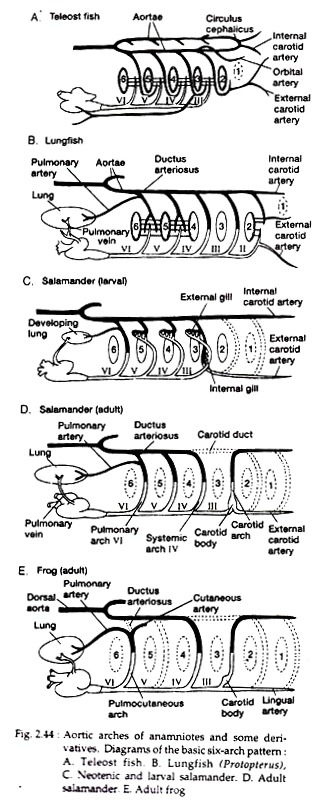
The remaining aortic arches (II – VI) complete the circle (Fig. 2.44A). The external carotid artery arises embryo-logically from the anterior end of the ventral aorta and becomes associated with the collecting loop, to carry oxygenated blood to the lower jaw.
The internal carotid artery supplying the brain, receives oxygenated blood from the first fully functional collecting loop (pharyngeal slit II) via the efferent branchial artery (II) (Fig. 2.44B).
Modifications in Amphibians:
ADVERTISEMENTS:
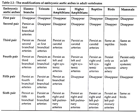
In amphibians, the first two aortic arches (I and II) disappear early in the development. The distribution pattern of other arches differs between larvae and metamorphosed adults. In most larval salamanders, the IIIrd to Vth aortic arches carry blood to the external gills, and the last aortic arch (VI) sprouts the pulmonary artery to the developing lung (Fig. 2.44C). During metamorphosis, the external gills are lost, but the aortic arches are retained as major systemic vessels.
The short section of dorsal aorta between aortic arches III and IV, termed the carotid duct, usually closes at metamorphosis. The section of neutral aorta between arches III and IV becomes the common carotid artery, which supplies the external and internal carotids.
The carotid body is an enlarged portion of the carotid artery that usually forms near the point at which the common carotids branch. Carotid body plays a role in sensing the gas content or pressure of the blood and may have some endocrine functions (Fig. 2.44D).
ADVERTISEMENTS:
The IV and Vth aortic arches constitute major systemic vessels that join the dorsal aorta. The Vlth aortic arch also joins the dorsal aorta, its last short section forming the ductus arteriosus. Shortly before joining the dorsal aorta, it gives off the pulmonary artery, which again divides into small branches to the floor of the mouth, pharynx, and oesophagus before actually entering the lungs.
In frogs, the larva has internal gills that reside on the last four aortic arches (III-VI), and the embryonic pulmonary artery comes out from Vlth arch. During metamorphosis internal gills are lost together with the carotid duct and all of arch V.
The III, IV and VI arotic arches persist and expand to supply blood to the head, body and pulmonary circuits, respectively (Fig. 2.44E). The IIIrd arch becomes internal carotid. The anterior extension of ventral aorta becomes external carotid. The Vlth arch loses its connection to the dorsal aorta because the ductus arteriosus closes and becomes the pulmocutaneous artery.
Modifications in Reptiles:
The symmetrical nature of aortic arches of the embryo tends to become asymmetrical in adult. This feature appears first in reptiles and then birds and mammals. The III, IV and VI aortic arches persist in reptiles. Maximum modification occurs in arch IV.
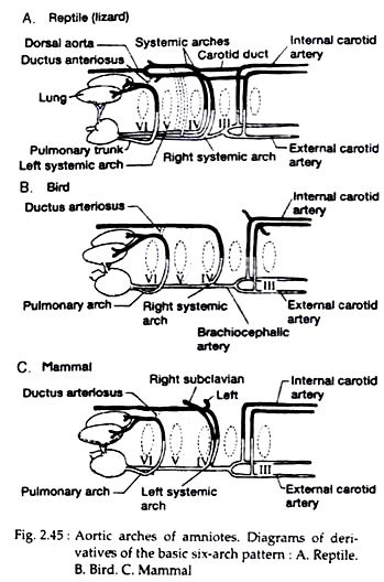
During embryonic development, the ventral aorta splits to form the bases of three separate arteries leaving the heart — the left aortic arch, the right aortic arch and the pulmonary trunk (Fig. 2.45A).
The left aortic arch (IV) and the curved section of the left dorsal aorta constitute the left systemic arch. The right systemic arch includes the same components of the right side of the body. The two systemic arches unite behind the heart to form the common dorsal aorta. The right systemic arch gives rise to many additional vessels than left systemic arch.
Modifications in Birds:
Only right systemic arch develops in birds. The bases of the right aortic arch (IV) and the adjoining section of the right dorsal aorta form the right systemic arch during embryonic development. The right aortic arch III and parts of the neutral and dorsal aortae contribute to form the carotids (Fig. 2.45B).
Modifications in Mammals:
Only three embryonic aortic arches persist in the adult mammals — the carotid, pulmonary and systemic arch (Fig. 2.45C). Carotid and pulmonary arteries develop from same components as in case of reptiles.
ADVERTISEMENTS:
The systemic arch arises embryo logically from the left IV aortic arch and left member of the paired dorsal aorta and therefore, is a left systemic arch in mammals. Development of subclavian arteries is peculiar — the left subclavian departs from the left systemic arch, while the right subclavian includes the right aortic arch (IV), part of the adjoining right dorsal aorta.
Portal system in vertebrates:
Portal system is the part of the venous system. A portal vein has its origin in capillaries and it ends in capillaries. The blood from the portal vein returns to heart through an intermediate organ. There are two types of portal systems in vertebrates – hepatic portal system and renal portal system. In hepatic portal system, capillaries from the gut unite to form hepatic portal veins, which again breaks up into capillaries in liver.
In case of renal portal system, the capillaries from the posterior part of the body unite to form renal portal veins, which, in turn, break-up into capillaries in the kidneys on their way to heart. The renal portal system is present in all classes of vertebrates except mammals. Mammals have only hepatic portal system.
Functions of portal systems:
The hepatic portal vein transports absorbed nutrients directly from the digestive tract to the liver for storage or processing of many end products of digestion. The renal portal system provides a direct route for delivering metabolic by-products to the kidneys that result from active locomotion involving the caudal musculature. The renal portal system also provides a way of improving kidney filtration.
Renal arteries entering directly from dorsal aorta to kidney have high pressure. The venous blood of the renal portal system has low pressure. Kidney filtration depends in part on an initial high pressure to move fluid out of the blood and into kidney tubules. But low pressure in the renal portal system aids in the recovery of water and other usable solutes and returning these fluids to the general circulation.


