In vertebrates the skin may be respiratory (e.g., anurans), while in some fishes and aquatic turtles, the vascular rectum or cloaca is respiratory. But there are two main types of respiratory organs- gills for aquatic respiration and lungs for aerial respiration. Both gills and lungs may occur in the same animal.
Accessory respiratory organs are also present in some vertebrates. In both kinds of respiration two conditions are essential; firstly the respiratory organs must have a rich blood supply with very thin moist epithelium covering the blood vessels so that these blood vessels are through into close contact with the environment (water or air).
Secondly in the organs of respiration the blood vessels should be reduced to thin capillaries which expose a large surface area to the environment, so that blood is brought into close contact with the water or air in the respiratory organs.
Exchange of oxygen and carbon dioxide occurs at two places, i.e., in the respiratory organs and in tissues. During internal respiration or tissue respiration exchange of O2 and CO2 occurs between blood and tissues (cells) of the body. During external respiration, gaseous exchange takes place between blood and external environment (e.g., in aerial respiration within lungs and in aquatic respiration within water and gills surface).
ADVERTISEMENTS:
In lower aquatic vertebrates the respiratory organs are not connected to the olfactory organs, but in air-breathing vertebrates there is a close association between the two. In Choanichthyes there is a direct connection between the olfactory and respiratory organs in which the internal nares or choanae open from the nasal cavities into the buccal cavity, but it is only in tetrapoda that air enters through the nasal cavities into the buccal cavity and then into the lungs.
Gills:
Gills are used for aquatic respiration found in fishes and amphibians. Gills are lacking in anamniotes. Besides exchange of gases at the surface of gills, salts are also eliminated from the gills surface in marine teleosts. Gills are of two types on the basis of their position- external gills and internal gills.
1. External Gills:
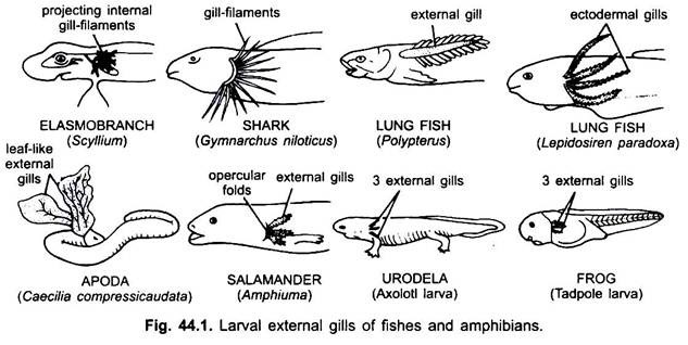
In larvae of most amphibians the integument covering the outer surface of visceral arches gives off branching outgrowths which are tufts of filaments and are respiratory. Thus, these are of ectodermal origin. In some tailed amphibians, external gills and gill-slits are retained throughout life, but in some tailed and all tailless amphibians they are lost during metamorphosis and, hence, called larval gills.
ADVERTISEMENTS:
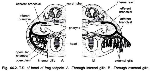
In the embryo usually five pairs of pharyngeal pouches arise, of which only second, third, and, fourth perforate and open to the exterior. In most amphibians these gill-clefts are closed during metamorphosis. The third, fourth, and fifth visceral arches bear external gills. External gills are in direct contact with water and an exchange of gases occurs through their surface epithelium.
Later an operculum arises and covers these gill- clefts and gills externally in tadpoles so that the gills become enclosed in an opercular chamber lined with ectoderm. These external gills soon degenerate and a new set of gills, called internal gills develop from the same visceral arches. However, these internal gills and operculum are not homologous with those of fishes.
External gills, though rare in fishes, are found in some larval forms of lampreys, Polypterus (bichir) has one pair of external gills. Dipnoi (Lepidosireri) have four pairs of filamentous external gills attached to the outer edges of the branchial arches.
The external gills of fishes disappear in the adult. Gills may be pectinate, bipinnate, dendritic or leaf-like. A gill has a narrow central axis bearing double row of filaments. These are richly vascularised by aortic arches. Gill-slits are not found.
2. Internal or True Gills:
Internal gills are characteristic of fishes. These are placed in the gill-slits and attached to the visceral arches. In amniotes embryonic pharyngeal pouches do not communicate to the outside through gill-slits in the adults. Gills are not found in them.
Gill-Slits:
Embryologically gill-cleft develops as a result of a series of evaginations from the pharynx which grow outward and meet corresponding ectodermal invaginations from the outside. At each point of junction a cleft or pouch is formed so that water coming into the pharynx from the mouth may pass to outside.
The partitions between these passage ways contain cartilaginous or bony supports for the gill-filaments which are located on each side. These gill-slits persist in the adults of cyclostomes, fishes and certain amphibians, but become abolished in higher vertebrates. The number of gill-slits is 6-14 pairs in cyclostomes, 5 to 7 pairs in sharks and rays, 4 pairs in most bony fishes.
Structure of a True Gill:
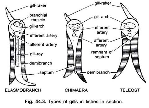
These gills develop on the walls of gill-pouches or gill-arches. A gill is formed of two rows of a series of gill-filaments or gill-lamellae develop from the epithelium covering the interbranchial septum on both sides. Interbranchial septum extends outward from the cartilaginous arch.
A single row of gill-filaments on one side of interbranchial septum forms a half gill called a hemibranch or demibranch. An interbranchial septum with two hemibranchs (anterior and posterior) forms a complete gill or holobranch. These are richly supplied with blood vessels associated with aortic arches so that carbon dioxide in the blood may be exchanged for dissolved oxygen in the water.
ADVERTISEMENTS:
Sharks and rays have generalised structure of gills. In higher bony fishes the interbranchial septum is lacking so that the hemibranchs on the anterior and posterior part of each branchial arch are no longer separated from one another.
Furthermore, the gill-apertures also no longer open separately to the outside. Instead, the gills are enclosed in a single chamber and covered externally by a large bony operculum which opens and closes posteriorly to permit water to pass to the outside. In most bony fishes there are four pairs of functional gills.
Spiracles:
In sharks and rays an anterior pair of non-respiratory openings, one on either side between mandibular and hyoid arches are called spiracles or pseudobranchs. These are internally closed. These become closed or lost in lung fishes and bony fishes.
Lungs:
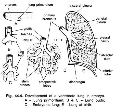
Most adult amphibians and all amniotes breathe by means of lungs, though lungs are also present in lung fishes. In an embryo a hollow outpushing, called lung primordium arises from the ventral wall of the pharynx. It grows backwards and divides into two, right and left lung buds. The undivided proximal portion develops into trachea and larynx, and opens into pharynx by glottis.
Later lung buds grow posteriorly into coelom and branch repeatedly and get covered by mesoderm. Thus, each lung has an endodermal lining and an outer visceral peritoneum and in between the two mesodermal mesenchyme having blood and lymph vessels, nerves, and smooth muscle fibres and connective tissue. Inner endodermal epithelium of lungs is raised into a network of ridges to increase the vascularised surface exposed to the action of air.
In lower forms, the lungs are hollow bags, but in higher forms the ridges increase in number and unite with one another across the lumen of the lung to convert it into a solid but spongy structure with innumerable air spaces. In mammals, the internal surface area of lungs may be thirty times that of the external surface area of the body.
The original duct of the lung sac connecting the pharynx to the lungs becomes a trachea in most. Trachea is absent in anurans. In many tetrapoda the anterior end of the trachea becomes modified into a larynx or sound box which opens into the pharynx by a glottis. At its lower end, the trachea divides into two bronchi, each of which enters a lung.
The bronchi divide to form an immense system of bronchioles carrying air into minute bags or alveoli. The alveoli have very thin walls invested with blood capillaries, an exchange of gases occur in the alveoli.
Larynx:
The upper end of trachea is enlarged, especially in frogs and toads, to form the larynx or sound box in which the vocal cords are located. In Necturus, it is supported by a pair of lateral cartilages bounding the glottis. In other amphibians, each lateral cartilage is divided into a dorsal arytenoid and a ventral cricoid cartilage.
In frog, both the cricoid fuse to form a cartilaginous ring. Larynx is not more developed in reptiles. Larynx is not sound producing organ in birds, but serves to modulate tones that originate in the syrinx. Syrinx lies at the lower end of trachea where it divides into two bronchi.
It is the sound producing organ. Larynx is greatly developed in mammals. Its wall is supported by a pair of arytenoid, single cricoid and a single thyroid cartilage on the ventral surface. Glottis may be closed at the time of swallowing of food by a flap of muscular epiglottis.
Trachea:
Trachea is extremely short or absent in Anura. It is merged with the larynx to form laryngotracheal chamber. Many caudate amphibians possess a short trachea, supported by cartilages. Trachea is simple in reptiles as in amphibians or may be long in long-necked reptiles such as turtles, trachea is long and convoluted.
Tracheal cartilages are sometimes in the form of complete rings. In birds, the trachea is long. In swans and cranes, trachea is longer than the neck and tracheal rings are complete and ossified. Trachea in mammals is variable and tracheal rings are usually incomplete on the upper side.
Lungs:
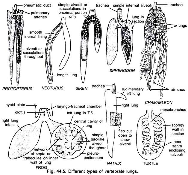
In Polypterus (African bichir) paired ventral lungs are present which enable these to survive during periods of draught. Dipnoans belonging to subclass Sarcopterygii which branched off from Actinopterygii also have a lung-like structure. In all the living lung fishes, the lung is dorsal to the gut connected by a tube to the ventral side of the oesophagus.
In Protopterus (African) and South American Lepidosiren it is bilobed and unpaired in Australian lung fish. Their lungs unlike Polypterus contain internal chambers or pockets to increase the respiratory surface and highly vascularised by branches of pulmonary arteries and veins. In most bony fishes and presumably the crossopterygians also, the primitive lung has modified into a gas or swim bladder or hydrostatic organ.
It may or may not be connected with the oesophagus by a dorsal connection. In amphibians the lungs are simple, sac-like structures with a central large cavity. In aquatic amphibians the inner surface of lungs is smooth. In frogs and toads the inner walls contain numerous folds lined with alveoli so as to increase the respiratory surface. They are richly vascular and lined with mucous epithelium whose cells are columnar and ciliated.
In reptiles, lungs are more complex than those of amphibians with an increase in the number of internal chambers and alveoli. In some lizards one lung is considerably larger than the other, and in snakes the left lung is reduced or even absent in some species. Crocodilians possess lungs that are quite similar to those of mammals. A few lizards possess diverticula, extending posteriorly from the lungs, resembling air sacs of birds. In some lizards, the bronchi are subdivided into primary, secondary and tertiary bronchi.
In birds, the lungs are small and incapable of the great amount of expansion. The lungs, however, are connected with nine air sacs that are situated in various parts of the body. The air sacs have no respiratory epithelium, serve essentially as reservoirs. Air passes through the bronchial circuit into the air sacs and then returns, generally by a separate set of bronchi, to the air capillaries in the lungs.
The respiratory system of the mammal is much less complicated than that of the bird. The primary bronchi after entering the lung into secondary bronchi which divide into smaller and smaller bronchioles, finally terminating in tiny alveoli or blind pockets in which there is an exchange of gases.
In most mammals, lungs are subdivided externally into lobes, i.e., left lung has two lobes and right lung has three lobes in man and four lobes in rabbit. Lungs are simple and without lobes in whales, sirenians, elephants, hyrax and several perissodactyles. Right lung is lobulated in monotremes and rats. In sirenians, the lungs are elongated.
Mechanism of Respiration:
In fishes and amphibians the mechanism of respiration is the same, the floor of the buccal cavity is lowered and water (fishes) or air (amphibians) is taken in, then the mouth is closed and the floor of the buccal cavity is raised which forces the water into gill-clefts in fishes or the air into the lungs in amphibians.
In amniotes air is taken in by increasing the volume of the lungs by an expansion of the thorax, this is done by movement of the ribs (and by movements of the diaphragm in mammals). In turtles where ribs are fixed to the carapace, the volume of lungs is increased by movements of the neck and limbs.
Accessory Respiratory Organs:
Gills are the chief respiratory organs in aquatic vertebrates, like fishes and some aquatic urodeles, etc. The land vertebrates have the lungs for respiration. There are also other accessory structures for respiration, i.e., for taking oxygen directly from water or air.
1. Skin:
Some fishes are able to survive outside water. The common eel, Anguilla can travel by wriggling on damp grass though it has no special respiratory organs, but it has vascular areas in the skin by which it can breathe both in water and on land. Secondly the opening of the operculum is small and rounded so that the eel can retain water in the branchial chamber and journey on land.
In amphibians also, the moist skin is highly vascular. Lungless salamanders (plethodonts) respire only through skin. Their larvae loses gills at metamorphosis and lungs do not develop in adults. African male hairy frog, Astylosternus have vascular hairy cutaneous outgrowths which act as respiratory surface.
Vascular caudal fin of Periophthalmus (mud-skipper) acts as respiratory organ during submergence. In the mud-skipper Periophthalmus of the Indian and Pacific Oceans the caudal fin is highly vascular, the head and trunk of the fish project above water when it perches on a rock, only the caudal fin remains submerged and acts as a respiratory organ.
2. Swim-Bladders:
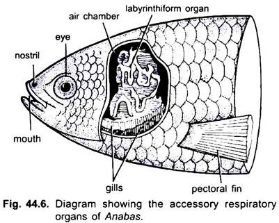
(i) The Indian climbing perch Anabas scandens (Fig. 44.6) has special air chambers above the gills, where three concentrically folded bony laminae, called labyrinth form organs are developed from the first epibranchial bone on each side. Their covering vascular mucous membrane brings about respiration. Anbas is so dependent on air that even in water it comes to the surface to gulp air and it is asphyxiated if prevented from doing so. It can survive for long periods on land and makes excursions by means of its many long spines on the operculum and ventral fins.
(ii) In Ophiocephalus there is an accessory branchial cavity on each side above the gills.
3. Epithelial Lining:
The loach Misgurnus swallows air which passes through the intestine and is voided by the anus, the highly vascular mucous membrane absorbs oxygen from the air, and carbon dioxide is also passed through the anus.
There may be other special organs for gaseous interchange. In Calichthys rectal respiration takes place, the rectum is highly vascular into which water is alternately taken in and pumped out.
4. Pharyngeal Diverticula:
The Indian ‘Cuchia eel’ Amphipnous has poorly developed gills, but on each side of the body there is a vascular sac as an outgrowth of the pharynx which opens anteriorly into the first gill-cleft. These sacs are respiratory.
5. Branchial Diverticula:
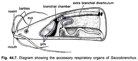
In the Indian catfish Saccobranchus (Fig. 44.7) there is a pair of large air sacs, each arising from the branchial chamber and extending laterally backwards into the trunk muscles. They can be filled with air for respiration.
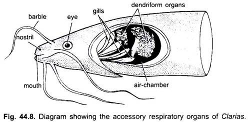
The catfish Clarias (Fig. 44.8), found in Indian and African rivers, has a pair of supra-branchial organs, each lying on one side and divided into two parts, a highly branched arborescent organ formed from second and fourth branchial arches, and a vascular sac of the branchial chamber which encloses the arborescent organ.
Several gill-fans formed by coalescing of gill-filaments close the entrance of the suprabranchial organ. Air is taken into the organ through the mouth continuously, and Clarias cannot only live outside water for several hours but it can move along on damp grass. Accessory respiratory organs are found generally in tropical fishes of amphibious habit, they are devices for sustaining life out of water.
6. Swim or Air-Bladders:
Swim or air-bladder arises as a diverticulum from the pharynx or oesophagus in bony fishes. It is originally lateral in position but becomes dorsal. It usually lies below or lateral to the vertebral column outside the coelom.
