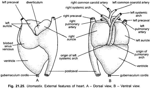
The blood vascular system of Uromastix is closed and consists of three main parts: 1. The Heart 2. The Blood Vessels (Arteries and Veins) 3. The Blood.
1. Heart:
The heart of Uromastix is typically reptilian and more advanced than the amphibians.
External Features:
It is a reddish coloured, triangular and muscular organ with its broad base forward or anterior and the pointed apex backward. It lies forward in the mid-ventral line in the pleuroperitoneal cavity below the sternum.
(i) Pericardium:
The heart is enclosed in a thin, firm, two-layered transparent sac, the pericardium, to which it is attached at its apex by a small cord, the gubernaculum cordis. It keeps the heart in position within the pericardium. The outer layer of pericardium is called the parietal pericardium, while the inner layer is termed visceral pericardium.
The parietal and visceral pericardium are continuous at the anterior end of the heart. Between the two layers of pericardium is a narrow space, the pericardial cavity, which contains a watery pericardial fluid. The pericardial fluid protects the heart from mechanical injuries and shocks and also allows it free movement.
(ii) Heart Chambers:
ADVERTISEMENTS:
The heart consists of four chambers: a large dorsal sinus venosus, two auricles (right and left) and a ventricle.
(a) Sinus Venosus:
The sinus venosus is a large thin- walled and bilobed chamber situated on the dorsal side of the heart covering both the auricles from above. It is formed by the union of a pair of precavals and a single postcaval veins. By a deep dorsal groove the sinus venosus is divided into two lobes, a larger right and a smaller left. The sinus venosus receives the deoxygenated blood from the body through the two precavals and a postcaval which open into the sinus by separate apertures.
(b) Auricles:
ADVERTISEMENTS:
There are two auricles- the right and the left. The two auricles are externally separated by a longitudinal interauricular groove. The right auricle is larger than the left. From its antero-dorsal edge, a small sac-like diverticulum is given off. This is said to be a normal feature of the heart in lizards. Its function is not known.
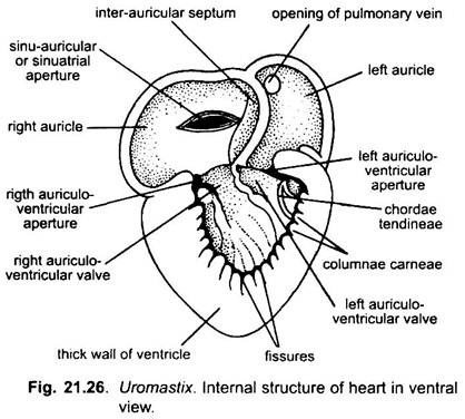
The internal lining of the right auricle is raised into numerous interlacing strands of muscular fibres called musculi pectinati. The right auricle opens into the lumen of the right side of the ventricle through the right side of the auriculo-ventricular opening which is guarded by the auriculo-ventricular valves. A small posterior inner margin of the right auricle extends into the lumen of the ventricle.
This portion of the right auricle which projects into the right side of the ventricle is called the atrium dextrum intraventriculare. The sinus venosus opens into the right auricle through a transversely elongated aperture called sinu-auricular aperture which is situated in the median dorsal wall of the right auricle. The sinu-auricular aperture is guarded by two lip-like sinu-auricular valves which project into the lumen of the right auricle.
According to Bhatia (1929), sinu-auricular valves are absent in Uromastix hardwikii. Only thick muscular lips keep the aperture ordinarily closed. It only opens when blood is forced through it into the right auricle.
(c) Ventricle:
The ventricle is a conical chamber whose walls are thick and spongy. It is separated from the auricles by a transverse auriculo-ventricular groove or coronary sulcus. Its apex is bluntly rounded and points a little to the right of the median axis of the heart and is attached to the pericardium by the gubernaculum cordis. The base of the ventricle is more or less straight. Its dorsal surface is slightly concave but the ventral is greatly convex.
Internal Structure:
It can be studied by cutting the section of heart.
ADVERTISEMENTS:
(i) Auricles:
The auricles are thin-walled. Both the auricles are separated from each other by a thin, muscular, unperforated and slightly curved partition, the inter-auricular septum. It is attached at its anterior end to the wall of auricles a little left of the diverticulum. At its posterior end the septum is free and reaches the lumen of the ventricle dividing the auriculo-ventricular aperture into right and left portions.
The left auricle is smaller than the right. It is also drawn out posteriorly into the lumen of the ventricle to form the atrium sinistrum intraventriculare. The pulmonary vein opens into the left auricle at its antero-dorsal surface, close to the inter-auricular septum, by a circular aperture not guarded by any valve.
(ii) Ventricle:
The thick and spongy walls of the ventricle are composed of a network of interlacing muscular fibres which greatly restrict the internal lumen. The inner surface of the ventricle is very uneven having numerous ridges and depressions. The ridges are called the columnae carneae and the depressions between them, the fissures. Auriculo-ventricular apertures of right and left sides are guarded by the flaps of the two auriculo- ventricular valves at its posterior end.
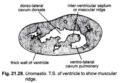
These two valves guard their respective portions of the auriculo- ventricular apertures. The free edges of the auriculo-ventricular valves are connected to the wall of the ventricle by firm cords called the chordae tendineae. A conspicuous flap, called the muscular ridge or interventricular septum, arises from the ventral wall of the ventricle on the left near the apex and extends horizontally forwards towards the right.
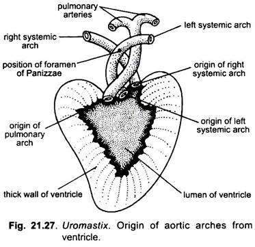
It divides the cavity of the ventricle into a large dorsal part, the cavum dorsale, and a small ventral part, the cavum pulmonale. The two parts keep communicating with one another over the margin of the ridge. Three aortic arches originate from the ventricle: a pulmonary arch and two systemic arches (right and left).
The pulmonary arch takes its origin from the cavum pulmonale, while the systemic arches arise from the cavum dorsale. Each of the three arches has a pair of semilunar valves at its base. The cavities of these valves face away from the ventricle to prevent the back-flow of blood.
Working of Heart:
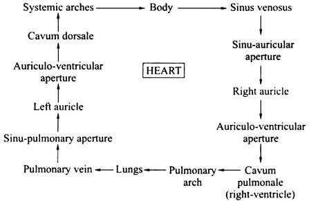
The sinus venosus receives deoxygenated blood from the whole body by way of three venae cavae. When the sinus venosus contracts, driving its blood into the right auricle through the sinu-auricular aperture. Meanwhile, the left auricle receives oxygenated blood from the lungs through the pulmonary vein.
Now the auricles contract almost simultaneously forcing the deoxygenated blood from the right auricle into the cavum pulmonale (right side of the ventricle). During this process, return of blood to the sinus venosus is prevented by the sinu-auricular valves. From the left auricle the oxygenated blood enters the cavum dorsale (left side of the ventricle).
In the middle portion of the ventricle, some mixing of pure and deoxygenated blood takes place. Now the ventricle contracts in such a way that the muscular ridge keeps the deoxygenated and oxygenated blood separate. The contraction of ventricle sends the deoxygenated blood from the cavum pulmonale into the pulmonary arch and the oxygenated and partly mixed blood from the cavum-dorsale into the systemic arches which is distributed to the entire body.
The head receives the purest blood. The return of blood from the ventricle into the auricles, during the contraction of ventricle, is prevented by the auriculo-ventricular valves. The return of blood from the aortic arches in the ventricle is checked by the semilunar valves at the base of arches. Therefore, it can be said that the heart of Uromastix is functionally four-chambered.
The blood circulation in the body is as follows:
2. Blood Vessels:
There are two types of blood vessels, viz., arteries and veins. The arteries carry blood from the heart to the various organs of the body and collectively form the arterial system. The veins collect blood from various organs of the body and return it to the heart and collectively form the venous system. The arteries and veins are connected by way of capillaries.
I. Arterial System:
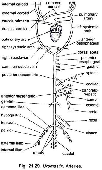
There are three aortic arches which arise independently from the ventricle, a little to the right of the middle line, viz., the pulmonary arch and a pair of systemic arches (right and left systemic arches).
A. Pulmonary Arch:
The pulmonary arch lies ventrally and arises from the right ventral side of the ventricle (cavum pulmonale). As it travels forwards, it curves and becomes dorsal to the left systemic arch, where it soon divides into right pulmonary and left pulmonary arteries going to the right and left lungs respectively.
B. Systemic Arches:
Both the systemic arches arise directly from the cavum dorsale of the ventricle carrying oxygenated blood. The left systemic arch is fully visible in the ventral view of the heart. It arises from the right side of the ventricle but curves later to the left and carries mixed blood. The point of origin of the right systemic arch is not visible from the ventral side but can be seen in the dorsal view.
The right systemic arch arises from the left side of the ventricle and lies dorsal to both the pulmonary and the left systemic arches. Later it curves to the right side. The two systemic arches communicate with one another by a foramen of Panizzae, situated where the arches cross each other anterior to the heart. Both the systemic arches then turn upwards, backwards and inwards and join posterior to the heart in the mid-dorsal line to form dorsal aorta.
The right systemic arch gives off a very short artery before it starts curving to the right in the notch between the two auricles which is called the carotis primaria or innominate artery. The right systemic arch also gives off a common subclavian artery before joining the left systemic arch.
(a) Common Carotid Arteries:
The innominate arises from the right systemic arch before curving to the right side. It bifurcates to form the right and left common carotid arteries. Each common carotid runs forward for some distance parallel to the systemic arch of its side and then divides into the external and the internal carotid arteries.
The external and the internal carotid arteries extend forwards through the neck, both of which branch into numerous arteries supplying oxygenated blood to the head anterior mesenteric region. Each internal carotid artery is connected with the systemic arch of its side by a vessel called the ductus caroticus.
It is a remnant of hypogastric embryonic lateral aorta. Since carotid arteries femoral arise from the right systemic arch, so they are also known as carotico-systemic arches. The ductus arteriosus or the ductus Botalli (which usually connects the pulmonary artery and the systemic arch) is absent in Uromastix hardwickii.
(b) Common Subclavian Artery:
It arises from the right systemic arch before it joins the left systemic arch. It soon divides into the right and the left subclavian arteries which run outwards to supply the respective forelimb. Each subclavian artery gives off three vessels, i.e., scapular to the scapular region, coracoid to the coracoid muscles and brachial to the arm.
(c) Anterior Oesophageal Artery:
The left systematic arch also gives off a small anterior oesophageal artery which supplies the anterior part of the oesophagus. Shah also described posterior oesophageal artery which arises from the dorsal aorta. However, Bhatia has figured only one oesophageal artery.
Dorsal aorta:
The dorsal aorta is formed by the union of the right and left systemic arches.
It extends straight backwards in the mid-dorsal line beneath the vertebral column, giving off the following arteries:
(i) Posterior Oesophageal Artery:
Immediately after the formation of the dorsal aorta a small unpaired artery arises from it which is called the posterior oesophageal artery. It supplies blood to the posterior part of the oesophagus.
(ii) Gastric Arteries:
Three small unpaired gastric arteries arise behind the posterior oesophageal artery. These arteries supply blood to the anterior, middle and posterior part of stomach and are called anterior, middle and posterior gastric arteries.
(iii) Posterior Mesenteric Artery:
The posterior mesenteric artery is large and unpaired, arises far forward at the level of the fifth parietal, just in front of the origin of coeliac. It runs backwards and distributes blood to the large intestine by three branches: the caecal, colic (colon) and rectal.
(iv) Coeliac Artery:
It arises behind the posterior mesenteric and runs forwards, crosses the posterior mesenteric and supplies blood to the spleen and to stomach by two branches, viz., the spleenic and gastric.
(v) Pancreo-Hepatic Artery:
It is a fairly large unpaired vessel supplying blood to the duodenum, liver and pancreas.
(vi) Anterior Mesenteric or Intestinal Artery:
It arises behind the pancreo-hepatic artery and supplies blood to the entire small intestine by a number of branches.
(vii) Genital or Gonadial Arteries:
These are paired, arise near the tenth parietal and supply blood to the gonads and suprarenal glands. They are called spermatic arteries in the male and ovarian arteries in the female.
(viii) Rectal Artery:
It is an unpaired artery, arises in between the thirteenth and fourteenth parietals. It passes down along the dorsal wall of the rectum and supplies blood to it.
(ix) Cloacal Artery:
It is an unpaired median artery which arises close to the common iliac arteries. It supplies blood to the lateral wall of the cloaca and the terminal portion of the rectum.
(x) Common Iliac Arteries:
A pair of large common iliac arteries arises near the middle region of the kidneys and enters the hindlimbs. Before entering the limbs, each common iliac artery gives off a hypogastric artery to the urinary bladder, a femoral artery to the thigh muscles and a pelvic artery to the pelvic girdle and the fat bodies. It supplies the hindlimbs by two branches, viz., external and internal iliac arteries.
(xi) Renal Arteries:
Three pairs of small renal arteries supply blood to the kidneys.
(xii) Caudal Artery:
The posterior end of the dorsal aorta continues behind as the caudal artery. The caudal artery runs through the haemal canal of the caudal vertebrae.
(xiii) Parietal Arteries:
During its course, the dorsal aorta also gives off about 15 pairs of small parietal arteries, supplying blood to the trunk muscles and caudal muscles.
II. Venous System:
The venous system of Uromastix is most primitive. The blood from the lungs is returned to the left auricle by two pulmonary veins. The blood from the other parts of the body is returned by three large veins.
The two precavals (right and left) return the blood from the anterior side, while the postcaval veins return the blood from the posterior side and enter into the bilobed sinus venosus which joins the right auricle.
Thus, the venous system of Uromastix has the following main veins:
a. Precaval veins;
b. Postcaval vein;
c. Hepatic portal vein;
d. Renal portal vein.
a. Precaval Veins:
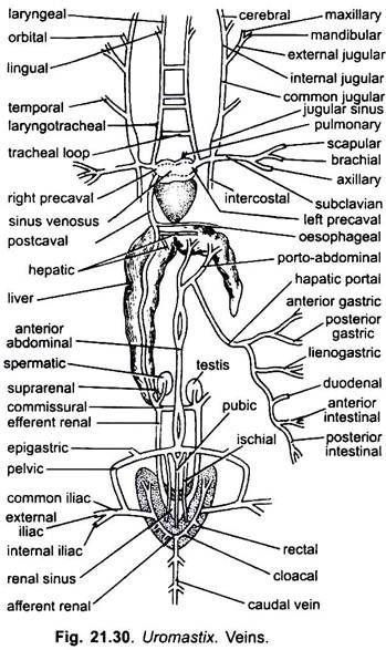
There are two precaval veins, the right and the left, which drain the blood from the anterior body region—head, neck, shoulders, forearms and thoracic wall.
Each precaval is formed by the union of four veins:
(i) Common jugular,
(ii) Subclavian,
(iii) Intercostal and
(iv) laryngo-tracheal.
(i) Common Jugular Vein:
The common jugular vein is formed by the union of external and internal jugular veins at the level of the tympanum. The external jugular vein receives blood from the upper jaw by a maxillary vein and from the lower jaw by a mandibular vein. The internal jugular receives blood from the brain by a cerebral vein and from the orbital region by an orbital vein.
The common jugular runs back into the neck where it receives blood from the auditory region of the head by a temporal vein. A little behind the junction of common jugular vein and temporal vein, there is a large sinus (expansion) called jugular sinus. Das (1960) has reported displacement of some elements of the external jugular.
This view is also confirmed by the embryological studies of Grossar and Brezina. However, some authors including Bhatia (1929) described that there is no external jugular on the left side and according to them the left precaval is formed by the union of an internal jugular and a subclavian only.
(ii) Subclavian Vein:
The large subclavian vein runs inwards from the forelimb and joins the posterior end of jugular sinus.
The subclavian vein receives blood from the arm through three small veins:
(a) Scapular from the shoulder region,
(b) Brachial from the arm, and
(c) Axillary from the arm-pit and thorax.
(iii) Intercostal Vein:
The intercostal vein collects blood from the skin and muscles of the chest and runs forwards to meet the precaval vein internal to subclavian. By the union of common jugular, temporal, subclavian and intercostal, a large precaval vein is formed that opens into the sinus venosus.
(iv) Laryngo-Tracheal Vein:
The laryngo-tracheal vein is a very slender vessel. It receives a very fine lingual vein from the tongue and a laryngeal vein from the larynx and trachea. It then passes backwards alongside of the trachea and joins the precaval close to its entry into the sinus venosus.
The two laryngo-tracheal veins are symmetrical running parallel to the trachea and are interconnected by four or five transverse tracheal loops. Bhatia (1929) described that the left laryngo-tracheal vein does not unite with the left precaval.
b. Postcaval Vein:
The postcaval vein opens into the right posterior angle of the sinus venosus. It collects blood from the posterior region of the body, i.e., kidneys, gonads and liver. It arises from the region of kidneys. Blood from the kidneys is collected through capillaries in a thin-walled renal sinus lying in the middle region of the kidneys.
A pair of efferent renal veins commences from the renal sinus and pass forwards along the sides of the vertebral column up to the gonads, being separated from one another by a pad of connective tissue. The right efferent renal vein is thicker than the left. Both the efferent renal veins receive blood from dorsal bodywall through a few small veins in the way.
Just behind the gonad, the left efferent renal vein sharply bends medially and joins the right efferent renal vein to form the commissural vein. A small genital vein from the gonads and a fine supra-renal vein from the supra-renal gland on each side pours into the commissural vein or the efferent renal vein of that side.
Only the right efferent renal vein proceeds as the postcaval vein, which passes through the right lobe of the liver and opens into the sinus venosus. The postcaval vein during its course also receives two or more hepatic veins from the liver and a small vertebral vein from the backbone. A small oesophageal vein from the oesophagus joins the postcaval after its emergence from the liver.
c. Pulmonary Vein:
There is a single pulmonary vein formed by the union of several veins from both the lungs. It runs along the ventro-lateral border of the trachea and passes dorsally to the sinus venosus before opening into the left auricle close to the inter-auricular septum. It is the only vein which carries oxygenated blood.
d. Hepatic Portal Vein:
Hepatic portal vein is a large vein which collects the blood from the alimentary canal by a number of fine branches and carries it to the liver. It is formed by the union of numerous veins from the different regions of the alimentary tract such as the anterior and posterior gastric veins from the stomach, lienogastric vein from the spleen and a part of the stomach, duodenal vein from the duodenum, anterior intestinal from the ileum and posterior intestinal from the caecum, colon, and rectum.
The hepatic portal vein and all its tributaries run in the mesentery. Anteriorly, the hepatic portal vein receives a branch of the anterior abdominal vein called the porto-abdominal vein and enters the middle of the left lobe of the liver where it capillarises. Blood from the liver as is collected by the hepatic veins and poured into the postcaval. The hepatic portal system is formed by the hepatic portal vein and porto-abdominal vein.
e. Renal Portal and Anterior Abdominal Veins:
The caudal vein starts from the tip of the tail and runs in the haemal canal of the caudal vertebrae receiving on its way several paired branches from the tail muscles. The caudal vein after reaching the kidneys divides into a pair of diverging renal portal or afferent renal veins.
The afferent renal veins pass over the ventral surface of the kidneys partly buried in their substance. They receive cloacal and rectal veins from the cloaca and rectum respectively. The renal portal vein of each side continues forward into the kidney, its side and ramifies into capillaries.
Blood from each hindlimb is collected by the internal (sciatic) and external iliac (femoral) veins. These two unite after entering the abdominal cavity to form a common iliac vein which is connected to the renal portal vein by a transverse connection (iliac-afferent union) near the middle of the kidney. Each common iliac vein extends forwards as the pelvic or lateral vein along the dorsal surface of the large yellow fat body.
The pelvic vein receives in the way fine adipose veins from the fat body, ischial vein and epigastric vein from the dorsal bodywall, vesicular vein from the urinary bladder and parietal veins from the posterior muscles. A fine median pubic vein joins both the pelvic veins. The two pelvic veins converge in front of the kidneys and unite in the mid-ventral line to form the anterior abdominal vein. The anterior abdominal vein is formed by the imperfect union of two pelvic veins.
In Uromastix, the anterior abdominal vein is double at places indicating a primitive feature of fishes where two lateral abdominal veins have not fused along their entire course. Anteriorly the two components of the anterior abdominal vein remain separate, one of the anterior abdominal vein joins with the hepatic portal vein and enters the left lobe of the liver as porto-abdominal vein, while another enters the left lobe of liver directly.
3. Blood:
Uromastix (reptiles) are cold-blooded or poikilothermous, like fishes and amphibians. Red cells are nucleated and survive for much longer than those of mammals. The total haemoglobin concentrations and oxygen carrying power of the blood are only half those of mammals. The white cells of reptiles include the same types as in other vertebrates.
