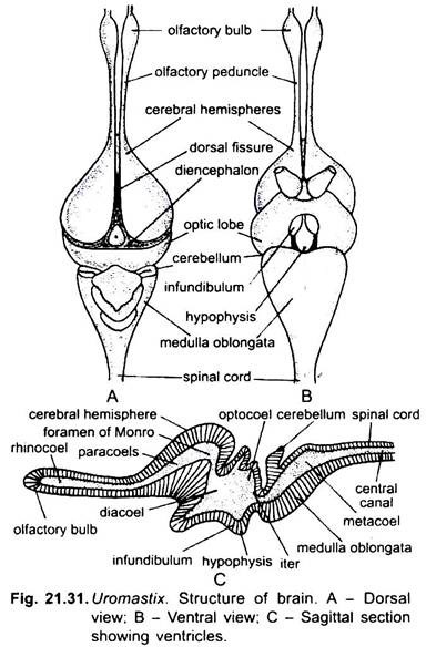
The nervous system in Uromastix is generally on the amphibian plan. Many differences in the plan of nervous system between the two groups are not apparent anatomically but reptiles, however, indicate a higher degree of nervous efficiency. One of the major differences is the absence of lateral line system in reptiles. The absence of this system has been fully compensated by improvements in the sensory organs.
Like other vertebrates, the nervous system of Uromastix consists of the following divisions:
A. Central nervous system includes the brain and spinal cord.
B. Peripheral nervous system includes the cranial and spinal nerves.
ADVERTISEMENTS:
C. Autonomic nervous system includes a pair of ganglionated lateral trunks.
A. Central Nervous System:
The central nervous system comprises the brain and spinal cord.
1. Brain:
In Uromastix, the brain is very simple and similar to that of the frog, however, it shows some advancement over that of the frog. The brain lies quite loosely in the cranial cavity lined with a tough fibrous membrane, duramater.
ADVERTISEMENTS:
Another membrane which surrounds the brain is called piamater. In between the two layers arachnoid fluid is present which protects the brain.
The brain of Uromastix is divisible into the three usual parts:
(i) The forebrain,
(ii) Midbrain and
ADVERTISEMENTS:
(iii) Hindbrain.
(i) Forebrain:
The forebrain forms the anterior part of the brain. It consists of large swollen and elongated cerebral hemispheres, olfactory bulbs and diencephalon.
(a) Cerebral Hemispheres:
The cerebral hemispheres are two oval bodies, somewhat narrow in front but broad behind closely applied together in the median line but a dorsal median longitudinal fissure forms the demarcation between the two.
(b) Olfactory Bulbs:
A pair of smaller olfactory bulbs and their posterior narrow stalks, the olfactory peduncles by which they are attached with the corresponding cerebral hemispheres, are the anteriormost parts of the forebrain. From olfactory bulbs originate the olfactory nerves. The cavity of each olfactory bulb is called rhinocoel.
ADVERTISEMENTS:
The roof of each cerebral hemisphere is thin and called the pallium and the thick ventral and lateral walls together constitute the corpus striatum. The pallium in Uromastix is, however, thicker than that of the amphibians. In Uromastix the cerebral hemispheres are large due to large fibrous tracts called corpora striata.
The two corpora striata are connected by a tranverse anterior commissure which receives many efferent fibres from the side-walls of the diencephalon. Above the anterior commissure lies the hippocampal commissure which connects the hippocampal regions of the cerebral hemispheres. Each cerebral hemisphere encloses a cavity, the lateral ventricle or paracoel which is continuous in front with the rhinocoel of the olfactory lobe of its side.
(c) Diencephalon:
The diencephalon is a small rounded area situated between the cerebral hemispheres and the midbrain. It is hardly visible from the dorsal side. The lateral walls of the diencephalon are thick forming the optic thalami. The thick floor forms the hypothalamus. The roof of the diencephalon is thin-walled and highly vascular forming the anterior choroid plexus.
Behind the anterior choroid plexus lie the habenular commissure and posterior commissure which is a transverse band of nerve fibres. From the thin dorsal surface of the diencephalon arises a median outgrowth, the pineal apparatus, which consists of two parts- the anterior parietal organ or parietal eye lying in the parietal foramen, and the posterior pineal body or epiphysis.
The parietal organ or parietal eye is considered to be functional eye. From the thick ventral surface of the diencephalon arises an infundibulum behind which is attached the pituitary body or hypophysis. Optic chiasma is situated anterior to the infundibulum.
Optic chiasma is formed by the crossing of two optic nerves. Diencephalon encloses a laterally compressed cavity, the diacoel or third ventricle, which communicates with the lateral ventricles of the cerebral hemispheres by a small common aperture, the foramen of Monro.
(ii) Midbrain:
The midbrain consists of two dorso-laterally placed rounded optic-lobes. On the ventral surface of the midbrain lie two bands of longitudinal, nerve fibres, stretching between the forebrain in front and the medulla oblongata behind, these are the crura cerebri.
Each optic lobe contains a ventricle, the optocoel. The two optocoels open into a narrow median canal, the iter, which connects the diacoel (cavity of diencephalon) in front and the metacoel (cavity of cerebellum) behind.
(iii) Hindbrain:
The hindbrain comprises the cerebellum and the medulla oblongata.
(a) Cerebellum:
The cerebellum is poorly developed as in the frog. It is merely a flattened semicircular ridge at the anterior dorsal surface of the medulla oblongata.
(b) Medulla Oblongata:
The medulla oblongata is the hindermost part of the brain. It is broad in front and narrow behind. It has a prominent ventral flexure where it passes into the spinal cord. The roof of the medulla is thin, vascular and folded forming the posterior choroid plexus. It encloses a cavity, the fourth ventricle or metacoel or myelocoel. Its cavity is connected with the diocoel through a narrow median passage, the iter.
All the ventricles of the brain are continuous and posteriorly connected with the cavity of the spinal cord and full of a watery fluid, the cerebro-spinal fluid secreted by the choroid plexuses.
The brain of Uromastix shows relative increase in size of the cerebral hemispheres. But the olfactory bulbs are reduced to a smaller size. The increase in the size of the cerebral hemispheres indicates an enlargement of instinctive activities. The behaviour of reptiles is certainly on a higher level than that of the amphibians in general. The reptiles respond more quicker than the amphibians.
Histology of Brain:
The brain is composed of two types of nervous tissue, viz., the grey matter and white matter. The grey matter is composed of nerve-cells and the white matter consists of nerve fibres. In the forebrain the grey matter is external and white matter is internal, while in the midbrain and medulla oblongata the arrangement is reverse of this.
Functions of Brain:
a. The olfactory lobes control the sense of smell.
b. The cerebral hemispheres get the impulses from the sense organs and initiate motor impulses for voluntary movements. The cerebral hemispheres are the seat of intelligence, memory, emotions and will.
c. The diencephalon acts as a relay station in conveying the impulses to the cerebral hemispheres and as integrating centre for the autonomic nervous system,
d. The optic lobes are concerned with the sense of sight. The crura cerebri carry the sensory impulses from the medulla oblongata and the spinal cord to the optic thalami and thence to the cerebral hemispheres.
e. The cerebellum coordinates the voluntary movements and also maintains the equilibrium.
f. The medulla oblongata controls the involuntary movements. Therefore, it is regarded as the most important part of the brain.
2. Spinal Cord:
The spinal cord is a long, whitish, somewhat dorso-ventrally flattened tube. It is situated in the neural canal of the vertebral column. It is also covered by the same meanings which cover the brain. A very shallow groove, the dorsal fissure, occurs along the mid-dorsal line of the cord. A prominent mid-ventral groove called the ventral fissure extends along the entire length of the spinal cord.
A thin longitudinal vertical septum of connective tissue lies in the thickness of the spinal cord beneath the dorsal fissure. The spinal cord encloses a narrow central cavity known as central canal. The central canal opens anteriorly into the fourth ventricle of the medulla oblongata but is closed posteriorly. It is filled with the cerebro-spinal fluid.
The spinal cord is composed of two types of nervous tissue- the inner grey matter and the outer white matter. In section the grey matter is H-shaped with prominent dorsal and ventral columns or cornua from which arise the dorsal and ventral roots of the spinal nerves. The grey matter is mainly composed of nerve cells and non-medullated nerve-fibres. The white matter is divisible into the dorsal, ventral and lateral funiculi. It comprises medullated nerve fibres.
The spinal cord conducts sensory and motor impulses to and from the brain. It is also a centre of reflex action.
B. Peripheral Nervous System:
The peripheral nervous system consists of twelve pairs of cranial nerves from the brain and sixteen pairs of spinal nerves from the spinal cord. The first ten pairs of the cranial nerves correspond with the ten pairs of cranial nerves of fishes and amphibians.
The eleventh or accessory and the twelfth or hypoglossal originate from the medulla behind the tenth pair and innervate the muscles of larynx, neck and tongue. The sixteen pairs of spinal nerves originate by two roots, dorsal and ventral, as in other vertebrates.
C. Autonomic Nervous System:
The autonomic nervous system consists of two conspicuous white ganglionated sympathetic trunks which are continued along each side of the vertebral column. Each of these autonomic trunk ganglion, is connected with the corresponding spinal nerve by ramus communicans. The autonomic nervous system performs both sensory and motor functions and helps to regulate involuntary reaction.
