The following description is of Lacerta. The external organs of taste are completely lacking in Reptilia. The sense of taste is confined to the mouth only.
Olfactory Organs:
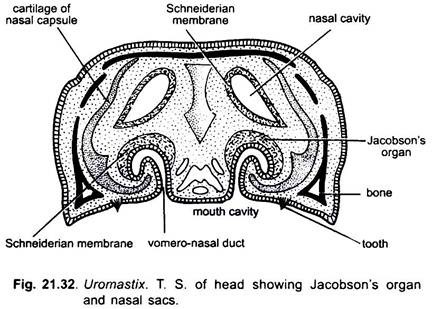
The olfactory organs are more complex than those of Anura. The nasal cavities open at the end of the snout by the external nares and into the cavity of the mouth by a pair of slit-like internal nares situated near the middle line of the palate. The external nare opens into the vestibule through which air passes to the sensory epithelium of the nasal or olfactory cavity proper. The nasal cavity contains a convoluted turbinal bone over which the mucous membrane extends.
Its cell are sensitive to chemicals in solution. The organs of chemical sense are better developed in reptiles. In reptiles, there are a pair of vomero-nasal organs or organs of Jacobson. The organs of Jacobson are well developed sac-like chambers lying below the nasal cavity but above the buccal cavity. Each organ consists of a blind sac lined with olfactory epithelium. It develops as a ventral hollow outgrowth of the nasal cavity. They are lined with olfactory epithelium, front of the internal nare.
Jacobson’s Organs:
ADVERTISEMENTS:
The organ of Jacobson is innervated by branches of the olfactory and the trigeminal nerves. This sense organ is important in most lizards and snakes and appreciates scent particles introduced into it by the tongue tips. These organs serve to smell the food when it has been taken into the buccal cavity. In some reptiles, they play a part in activities such as trailing prey and locating members of the opposite sex. Paired ducts communicate with the buccal cavity, probably enabling the olfactory appreciation of substances held in the mouth.
Eyes:
The eyes of Uromastix exhibit some advancement over those of the amphibians due to the transition from water to land. Although both the eyelids are movable but the lower eyelid is more movable in comparison to the upper eyelid. The eyelids close the eyes to protect them from the dust, heat and rain. There is a third eyelid, the nictitating membrane, in the anterior comer of the eye which develops and lies folded beneath the lower eyelid.
It is really a double fold of conjunctiva and is not homologous with that of the frog. The nictitating membrane can be drawn rapidly over the moist cornea and, thus, it protects and cleans the eye. There are glands which keep the eyelids and cornea moist and clean.
The Harderian gland lying on the anterior side of the eyeball lubricates the nictitating membrane, and the lacrymal gland lying on the posterior side of the eyelid keeps the eye moist and clean. The tears (water fluid) are passed from the eye into the nose through a lacrymal duct.
ADVERTISEMENTS:
The eye of Uromastix consists of the usual three layers, viz.,
(i) The outer fibrous tunic or sclerotic,
(ii) The middle uvea or choroid and
(iii) The inner retina.
ADVERTISEMENTS:
(i) Fibrous Tunic:
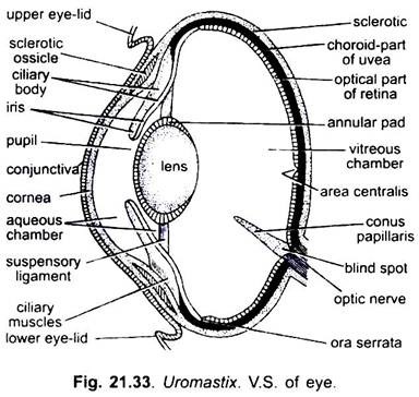
The thick and tough fibrous tunic protects the eyeball and maintains its form. The fibrous tunic is distinguished into two distinct regions- an anterior, small, transparent and exposed portion called the cornea and a posterior, large, opaque cartilaginous portion called the sclerotic lying hidden in the orbit. A small anterior portion of the sclerotic, the cornea is, however, visible and is commonly called the white of the eye.
The cornea is supported by a ring of eleven small sclerotic bones, or ossicles, present around the iris. The cornea is curved and provides the main refracting surface. A thin transparent epithelial layer is fused to the outer surface of the cornea. It is known as conjunctiva. The conjunctiva is continued over the inner surface of the eyelids.
(ii) Uvea:
Uvea is differentiated into three regions:
(a) A greater part of it is thin, vascular and pigmented and lies in close contact with the sclerotic. It is called the choroid. It serves to darken the eye.
(b) At the junction of the sclerotic and cornea, the uvea forms a thick ring which constitutes a part of the ciliary body,
(c) Just in front of the cavity of the eye, forming a part of the circular partition is the iris. The iris is perforated at the centre by an aperture, the pupil. The iris possesses circular and radial muscles to reduce and enlarge the pupil respectively. The iris contains pigment cells, which impart it its characteristic light-orange colour.
(iii) Retina:
ADVERTISEMENTS:
Retina lies inside the entire uvea and is differentiated into distinct regions:
(a) Its portion lying in contact with the choroid is thick and sensitive and is called the optical part of retina. It contains more cones than rods, thus, there is a good daylight vision and probably good colour perception.
This condition is found in most lizards (diurnal types) and many turtles. The double cones of turtles and lizards may serve to detect polarized light. This portion is protected from excessive exposure to light by yellow droplets found over this portion,
(b) Remaining retina is thin and non-sensory. It lines the iris and ciliary body. The part forming the ciliary body is known as the ciliary part and that to the iris as the iridial part. The junction of the optical and ciliary parts of the retina is irregular and is called ora serrata.
The lens is a biconvex, transparent body lying immediately behind the iris. It is more convex behind than in front. A ring of soft tissue, the annular pad, surrounds the circumference of the lens. The lens is suspended and held in position by radially arranged fibres which arise from the ciliary body and are attached to the annular pad and form the suspensory ligament. The suspensory ligament is formed of ciliary muscles and ciliary processes.
The lens and its suspensory ligament divide the cavity of the eye into two unequal compartments, viz., the anterior small aqueous chamber and the posterior large vitreous chamber. The aqueous chamber is further divided by iris into anterior and posterior parts containing a clear watery fluid, the aqueous humour, and are continuous through the pupil. The aqueous humour keeps the eyeball taut or stretched for distinct vision.
The optic nerve arises from the inner surface of the retina and joins the brain after piercing through all the three eye-coats at the back of the eyeball. The point of origin of optic nerve is called the blind spot. No image is formed at this point. A small part of the retina opposite the centre of the lens is the place of the most distinct vision. It is called the area centralis or yellow spot. A peculiar pigmented, blackish brown, highly vascular, cushion-like rod, the conns papillaris, projects into the vitreous humour from the blind spot. Probably it provides nutrition, like the pecten of birds.
Accommodation for near vision is usually produced by the striated ciliary muscles, so arranged that they cause the ciliary process to squeeze the lens, making its anterior surface more rounded.
Ear:
The ear of Uromastix consists of two principal parts- the middle ear and the internal ear. External ear is absent.
Middle Ear:
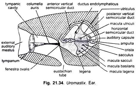
The middle ear comprises an air-filled cavity called the tympanic cavity. It is bounded externally by the tympanum and internally by the auditory capsule. The tympanus lies behind the jaws, sunk a little below the surface. From the lower part of the tympanic cavity a narrow passage, the Eustachian tube, extends downwards and inwards to open into the posterior part of the pharynx.
A slender rod-like ear ossicle, the columella auris, stretches across the tympanic cavity. The columella auris is divided into two parts: the inner bony stapes fitting into a small membrane covered aperture, the fenestra ovalis, and the outer cartilaginous extra stapes is attached to the inner surface of the tympanum.
Internal Ear:
The internal ear is situated in the auditory capsule. It is a soft, delicate and complicated structure known as membranous labyrinth or vestibule. Its upper cylindrical part is known as the utriculus and the lower large and rounded sacculus. Three semi-circular ducts (2 vertical and 1 horizontal) open into the utriculus at both of their ends. The two vertical ducts join together by their adjacent ends before opening into the utriculus. One end of each semicircular duct is enlarged to form an ampulla.
The sacculus gives rise to a small lobe, the pars lagena, which represents the cochlea of mammalian ear. It is an uncoiled tube and hearing is performed by it. Its wall, known as the limbus, is strengthened by a peculiar form of connective tissue. The receptor hairs (Organ of Corti) are carried on a basilar membrane and are in contact with a tectorial membrane attached to the limbus.
Hearing is good in some lizards which also produce sounds for communication. Snakes respond mainly to earth-borne vibrations through the quadrate attached to the stapes (tympanum is absent in snakes and some lizards also). A narrow ductus endolymphaticus extends dorsally from the saculus and ends in a small blind sac over the hindbrain.


