Quick Notes on Balanoglossus :- 1. Introduction to Balanoglossus 2. Food Collecting Organs in Balanoglossus: 3. Collection of Food 4. Filtration of Food.
Introduction to Balanoglossus:
Balanoglossus is an elongated worm-like burrowing animal in the sea shore and found all over the world. In the Gulf of Mannar and Sunderban region of India another hemichordate, Saccoglossus is prevalent. This animal excavates its own burrow in sand or sandy mud. The burrow is U-shaped with two openings, 10 to 20 cm apart. The anterior end of the burrow is funnel shaped.
During the time of high tide, the proboscis of Balanoglossus comes out of the burrow to explore the surface. The body is divisible into three major parts—proboscis, collar, and trunk.
Proboscis and collar help to collect food from sea water, while filtration and digestion take place in the digestive tract. These animals are ciliary feeders, collecting food particles from the outside through the agency of cilia and mucus, secreted by the body surface.
Food Collecting Organs in Balanoglossus:
ADVERTISEMENTS:
(a) Proboscis:
It is a short club-shaped structure, and forms the anterior most part of the body. The club shaped proboscis gradually narrows posteriorly to a proboscis stalk. The hollow proboscis communicates with the outside by a proboscis pore situated on its left side (Fig. 2.48). The epidermis of the proboscis is uniformly ciliated and contains a large number of secretory cells.
The secretory cells are mainly of two types — one is goblet shaped and the other is larger columnar to triangular shaped, having an appearance, ranging from granular to alveolar. The goblet cells secrete proteinaceous substances and the larger cells secrete acid mucopolysaccharides. Another type of cell, termed mulberry cell is reported in some species to secrete external amylase.
(b) Collar:
ADVERTISEMENTS:
The median part of the body consists of a flap of muscular tissue forming the collar. The proboscis stalk and a part of the posterior proboscis are covered by collar. The collar is hollow, having a paired cavity that opens to the exterior by two apertures.
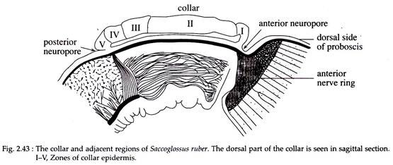
The collar houses the mouth on the ventral side. The collar epidermis is ciliated. According to the intensity of ciliation and characteristics of the secretory cells, the collar can be divided into five zones (Fig. 2.43). The first three zones (I, II & III) secrete acid mucopolysaccharides, while the last two zones (IV & V) secrete a protein rich material.
Collection of Food by Balanoglossus:
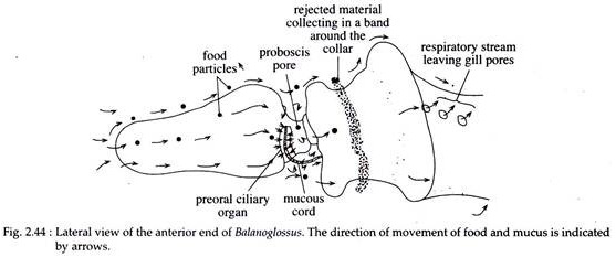
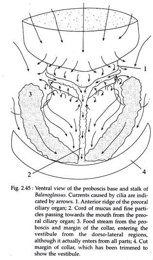
Balanoglossus collects food particles from the surrounding water. The particles from the substratum adhere to the mucus of the body surface and are carried by the continuous beat of cilia towards the mouth at the hinder end of the proboscis stalk (Fig. 2.45).
ADVERTISEMENTS:
At the base of the proboscis, the dorsal cilia beat lateroventrally. The mucus-entangled food particles forming threads are then directed on to the sides of the stalk and then towards the ventrally situated mouth (Fig. 2.44 and 2.45).
The rejections of unnecessary and unsuitable particles are made by the action of the muscular lip of the collar. The collar folds over the mouth so as to close it. The cilium of the collar transports the unwanted materials posteriorly.
Filtration of Food by Balanoglossus:
The food particles and mucus along with water come into the buccal cavity. From here it passes into the pharynx. The food particles are filtered from the water within the pharynx. Pharynx becomes specialized for this purpose. To understand the process, it is important to have a knowledge on the structure of pharynx.
Structure of Pharynx:
The pharynx is an elongated tubular structure, modified for respiration and food concentration. There are two distinct morphological divisions within the pharynx for performing two physiological functions. A para-pharyngeal ridge on each lateral wall divides the pharynx into two incomplete halves—a dorsal respiratory half pierced by gill slits and a ventral half that helps in food concentration.
The respiratory region of the pharynx is perforated by an longitudinal series of gill- slits. Each gill-slit is a U-shaped aperture. The portion of the pharyngeal wall separating two gill-slits is called gill bar. The gill bars are provided with strong ciliary armature. The gill-slits have no direct opening to the exterior. Each gill-slit leads into a pouch-like cavity called the branchial sac which in turn opens to the exterior by the gill pore.
The ventral food concentrating region of the pharynx has several longitudinal rows of long cilia interrupted by rows of cells with small cilia. Some secretory cells are present within the ciliated cells. The pharynx leads into the oesophagus.
ADVERTISEMENTS:
Mechanism of Food Concentration:
The mucus-entangled food particles are pushed into the mouth which reaches the pharynx through the buccal cavity. Most of the food material is directed to the ventral part of the pharynx as they are heavier in weight. The agent of the respiratory current that drives the water through the gill-slits are the outwardly directed beat of the strong lateral cilia of gill bars. These cilia beat in metachronal waves.
The finest food particles that float in the respiratory water, are collected by the synapticulae of the gill clefts and are transferred by ciliary action to the ventral region. Here, these particles are also entangled with the mucus. The ciliary beats of ventral region, pass the food material to the oesophagus, where digestion starts.
Digestion of Food:
Little is known about digestion in Balanoglossus. However, it has a distinct digestive tract.
Digestive Tract:
It is a straight tube starting from mouth to anus. The structure of pharynx has already been described. The pharynx leads into a short oesophagus which is followed posteriorly by the intestine. The intestine is a straight tube and is distinguishable into an anterior hepatic region, marked by sacculations of the dorsal wall of the intestine and a narrow post-hepatic region that opens to the exterior through the anus.
Digestive Process:
The peristaltic activity of the oesophagus, squeezes the food material through its lumen and compacts it ‘into a firm food cord. The well lubricated food cord moves posteriorly due to the beating of the ciliated digestive tract.
The enzymes of proboscis and oesophagus are responsible for digestion. A range of digestive enzymes are present, which can digest starch, glycogen, maltose, casein, sucrose etc. The digested material is absorbed in the intestine. Undigested material, together with sand particles and other debris are extruded from the burrow as characteristic castings.


