The below mentioned article provides an overview on Pheretima (Earthworm):- 1. Introduction to Pheretima 2. Habit and Habitat of Pheretima 3. Structure 4. Body Wall 5. Locomotion 6. Digestive System 7. Feeding and Digestion 8. Respiratory Phylum System 9. Circulatory System 10. Excretory System 11. Nervous System 12. Reproductive System.
Contents:
- Introduction to Pheretima
- Habit and Habitat of Pheretima
- Structure of Pheretima
- Body Wall of Pheretima
- Locomotion of Pheretima
- Digestive System of Pheretima
- Feeding and Digestion of Pheretima
- Respiratory Phylum System of Pheretima
- Circulatory System of Pheretima
- Excretory System of Pheretima
- Nervous System of Pheretima
- Reproductive System of Pheretima
1. Introduction to Pheretima:
Earthworms are common and very well known to most of us. The common earthworm of our country is known as Metaphire sp. It is also commonly found in Sri Lanka, Japan, Australia and South East Asia. It is represented by 13 species in the Indian soil. However, in this text we will describe Pheretima posthuma.
2. Habit and Habitat of Pheretima:
ADVERTISEMENTS:
Pheretima is a terrestrial earthworm, living in burrows made in moist soil. It prefers to live in burrow during daytime and at night and rainy season they come out of their shelter. It is thus nocturnal in habit. They are, however, absent in regions where the soil is sandy and deficient in humus.
The castings of earthworm are small rounded pellets or balls that lie at the opening of the burrow. These castings are formed when it goes deep into the hard and closely packed soil. The soil upon which it feeds, passes through the body and are deposited as castings. While burrowing, the earthworm makes the soil loose and porous.
The body wastes of earthworm increases the fertility of the soil. So, earthworms are often said to be the natural tillers of land. Vermiform culture or vermiculture is of much practice as earthworms are capable of converting kitchen wastes and other human wastes into fertilizers.
3. Structure of Pheretima:
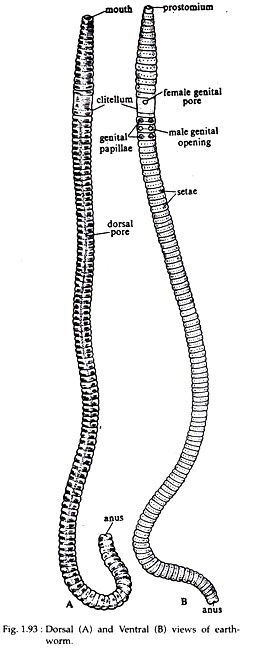
The body of earthworm is elongated, narrow and cylindrical (Fig. 1.93) measuring about 20 cm in length and 3 to 5 mm in width. The anterior end is more pointed than the posterior end. The dorsal side of the body is brown in colour and can be distinguished from the ventral side which is lighter in colour.
ADVERTISEMENTS:
The brown colour is due to the pigment porphyrin which is present in the body wall and it protects the body from bright and strong light. The impression of the dorsal blood vessel can be seen on the dorsal side as a dark median line extending throughout the length of the body.
The soft and naked body is made up of distinct segments or metameres separated from each other by inter-segmental ring-like grooves or annuli. The body is made up of a series of 100-120 similar segments. This external segmentation corresponds to internal segmentation and is referred to as metameric segmentation.
The segments 14 to 16 from the anterior end are encased in a thick glandular tissue sheet called the clitellum (saddle) or cingulum (belt) (Fig. 1.93). Considering the clitellum as the index, the body may be divided into three regions, namely the pre-clitellar, clitellar and post- clitellar regions. Some of the anterior segments bear superficial furrows and may appear to be subdivided, but these are merely external subdivisions.
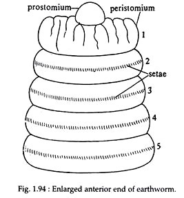
A distinct ‘head’ is absent in Pheretima. The first body segment is called peristomium (Greek : peri, around; stoma, mouth) which bears the mouth aperture on the ventral surface. The peristomium is prolonged anteriorly into a small, fleshy lobe, the prostomium (Greek: pro, anterior).
ADVERTISEMENTS:
The prostomium is thus considered as a projecting part of the first segment and not a segment by itself (Fig. 1.94). The last segment, at its posterior end bears the anus.
The body of earthworm consists of various apertures such as:
(i) Mouth:
It is a crescent-shaped aperture situated ventrally in the prostomium.
(ii) Anus:
It is a round aperture situated at the posterior end of the last segment.
(iii) Female Genital Aperture:
ADVERTISEMENTS:
The female genital aperture is a single opening situated on the mid-ventral line of the 14th segment, at the clitellar region.
(iv) Male Genital Apertures:
These are paired opening situated on the ventrolateral sides of the 18th segment, just below the clitellum. Each male genital aperture is associated with one pair of genital papillae, situated on the 17th and 19th segments (above and below the male genital aperture).
(v) Spermathecal Apertures:
The spermathecae opens to the outside through four pairs of small elliptical opening called spermathcal aperture. These are situated on the ventro-lateral side of the inter-segmental grooves between segments 5/6, 6/7, 7/8, 8/9. Through these apertures sperms are received from another earthworm during copulation.
(vi) Nephridiopores:
These are minute openings of the integumentary nephridia that open on the ventral surface of the body. They are present in large numbers, in all segments excepting the first six and the last one.
(vii) Dorsal Pores:
The dorsal pores lie as minute openings along the mid-dorsal line, one pore in each inter-segmental groove behind the 12th segment onwards, excepting the last one. The coelom communicates to the exterior by means of these pores. The coelomic fluid may be ejected outside to increase the surface moisture.
4. Body Wall of Pheretima:
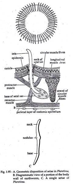
The body wall of earthworm is covered externally by a thin non-cellular cuticle (Fig. 1.95B) made up of parallel layers of collagenous fibres and is perforated by numerous pores through which open the epidermal glands.
Histologically, the cuticle comprises of two layers separated by an intervening layer. It is secreted by the supporting cells of the epidermis lying beneath it. Below the cuticle is the epidermis which is single layered.
The epidermis is made up of the following cells:
(i) Gland Cells or Goblet Cells:
These are mucus-secreting cells which keep the skin slimy and moist. Gland cells are of two types mucus cells and albumen cells.
(ii) Supporting Cells:
These are tall and columnar cells, forming the bulk of the epidermis and each has an oval nucleus in the middle.
(iii) Basal Cells:
They are small rounded or conical cells each with a distinct nucleus, lying between gland cells and supporting cells, which they can replace.
(iv) Sensory Cells:
They are usually cylindrical cells arranged in groups. Their outer ends give out minute hair-like processes which help to receive stimuli. Below the epidermis there is a circular muscle layer forming a thin continuous sheet around the body.
This is followed by a much thicker longitudinal muscle layer, running in parallel bundles, separated from one another by connective tissue and strengthened by collagen fibres. The muscle fibres are long, un-striped and spindle-shaped.
When the circular muscle fibres contract, the diameter of the body becomes narrow and the earthworm elongates. When the longitudinal muscle fibres contract, then earthworm becomes shorter and its diameter increases. The innermost layer of the body wall is formed by coelomic epithelium which is a thin membrane made up of single layer of cells (Fig. 1.95B).
Coelom:
The body cavity is a true coelom. It is extensive, but not continuous and is divided into a number of compartments by transverse partitions running between the body wall and the alimentary canal. These partitions or septa are vertical in disposition and are formed by double layers of peritoneum and numerous interlacing bundles of muscle fibres.
The septa are perforated by numerous sphinctered oval or circular apertures, through which communication is set up between adjacent coelomic chambers. The sphincter apertures are situated just dorsal to nerve cord in each septum except the first four segments.
In normal condition the sphincter muscles remain contracted keeping each coelomic compartment separated from each other. The coelom opens to the exterior by dorsal pores and nephridiopores.
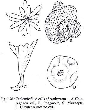
The number of coelomic compartments corresponds to the number of external segments, except the first four segments where septa are lacking. The coelomic compartments remain filled with a milky-white coelomic fluid. The coelomic fluid consists of plasma and four types of nucleated corpuscles (Fig. 1.96).
The four types of corpuscles are:
(i) Phagocytes:
These cells are saucer- shaped, granular, large and are most common, each having several folds on the surface.
(ii) Circular Nucleated Cells:
These are non-granular cells, nucleated and somewhat round in shape. They comprise of about 10% of the coelomic corpuscles.
(iii) Mucocytes:
These are elongated cells, each having a broad, fan-like process attached to a narrow nucleated body.
(iv) Chloragogen Cells:
These cells are small and are as numerous as the phagocytes. When stained with iodine solution, they become yellow. The body of each cell gives out numerous bulging’s.
5. Locomotion of Pheretima:
The locomotory organs of earthworm are the setae. Each seta is an elongated more or less ‘s’-shaped structure, composed of chitin, hardened and strengthened by the addition of sclerotised protein and is embedded in an epidermal pit called setigerous sac or setal sac. A seta measures about 0-24 mm in length and 0-03 mm in breadth.
The distal end of the seta is pointed and is called the neck. About one-third of the length of a seta is the neck and is the part that projects above the surface of the skin (Fig. 1.95B). The proximal end of the seta is blunt, called the base and it lies embedded in the skin. The middle of the seta is swollen and is called the nodulus (Fig. 1.95C).
The number of setae in each segment varies considerably. Usually they are numerous in each segment and are disposed in the form of a ring round each segment (Fig. 1.95A). Setae are present in all segments except the first, last and the clitellar segments.
Being built on a metameric plan, the locomotion of earthworm is the result of co-ordinated movements. When an earthworm starts to crawl, the first few segments become thinner and longer. This is caused by contraction of circular muscles and relaxation of longitudinal muscles of that region. The opposing sets of muscles are antagonized by an increase in the pressure of the coelomic fluid.
The thinning and elongation of the body gradually spread to more posterior segments. At this stage the setae of the anterior region are protruded to grip the substratum. The longitudinal muscles of the anterior region then contracts, so that the body at that region becomes shorter and stouter and the more hinder regions are pulled forward.
The septa also play an important role as they act as water-tight partitions which relay pressure changes from one segment to the next by bulging.
6. Digestive System of Pheretima:
The alimentary canal of earthworm is a long and straight tube of varying diameter and running from the anterior mouth to the posterior anus (Fig. 1.97). It is held in position by the inter segmental septa. The mouth is a crescent-shaped aperture situated ventrally on the peristomium. Mouth leads into a short, thin walled buccal cavity, which extends to the middle of the third segment and is surrounded by muscle strands.
A living earthworm can protrude and retract its buccal chamber, which acts as an. organ of ingestion of food. The buccal cavity leads into the pharynx lying in segments 3rd and 4th, and is internally marked off from the buccal chamber by a dorsal transverse groove. Pharynx is pear-shaped and its wall is thick and muscular. The dorsal wall of the pharynx is lobulated and richly vascular.
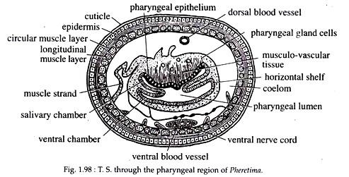
The lobulated part is called pharyngeal bulb containing saliva-secreting gland cells or chromophil cells, musculovascular tissue and ciliated epithelium. The lateral sides of the pharynx are pushed inside forming a narrow horizontal shelf on each side (Fig. 1.98). The two shelves meet anteriorly and posteriorly, dividing the pharyngeal cavity into a dorsal salivary chamber and a ventral conducting chamber.
From the pharyngeal wall running outwardly to the body wall are numerous radial dilatory muscles. The pharynx is followed by the oesophagus extending up to the 8th segment. Oesophagus is a straight, narrow, long and thin-walled tube.
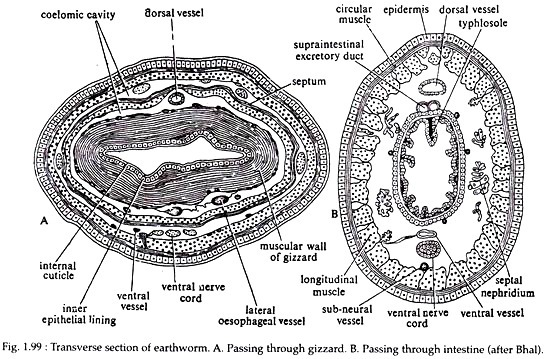
The part of the oesophagus lying in the 8th segment has become modified to form an oval structure called gizzard. The gizzard is oval, hard, thick-walled, muscular structure and its inner lining epithelium bears a distinct cuticle (Fig. 1.99A). The food particles are grinded against the cuticle into finer particles by the action of the muscles.
The part of the alimentary canal lying between segments 9 to 14 is called stomach. The wall of the stomach is highly glandular, vascular and thrown into internal folds. Both the ends of the stomach are provided with sphincter muscles. The region next to the stomach is the intestine, which is a long, wide and thin- walled tube, extending from the 15th to the last segment, up to the anus.
A pair of short and conical intestinal caeca is situated at the 26th segment. The dorsal wall of the intestine between the 26th and 95th segment is folded to form the typhlosole (Fig. 1.99B), so that in cross-section this part of the intestine appears to be ‘U’ shaped.
On the basis of the position of the typhlosole the intestine may be divided into three regions—pretyphlosolar region (from 15th to 26th segments), typhlosolar region (from 26th to 95th segments) and post-typhlosolar region (from 95th to last segments).
The role of the typhlosole is to increase the surface of absorption. Due to the presence of chloragogen cells in the lining of the intestine, the outer wall of it appears yellowish.
Histology of the Gut Wall:
The outermost layer of the gut wall is made up of tall and narrow cells derived from peritoneal epithelium. This layer often remains cohered by chloragogen cells, laden with yellow pigments. These yellow cells are believed to be excretory in function. Some workers believe that in addition, they also take up the role of digestive glands.
The peritoneal epithelium is followed by longitudinal muscle fibres and circular muscle fibres. The internal epithelium is formed of glandular and ciliated cells. A fourth layer in the form of cuticle is present only in the buccal cavity (thin layer) and in the gizzard (thick layer).
7. Feeding and Digestion of Pheretima:
Pheretima ingest soil, rich in inorganic particles, seeds, decaying leaves, ova and larvae of small animals. At night they come out of their holes to feed. The leaves or vegetable matters are seized by the pointed end of the mouth, the buccal chamber is everted and the food is drawn in by the suction of the pharynx.
Before the food is taken in, they are moistened by mucus secreted from the pharyngeal bulb. The food is forced to the oesophagus by peristalsis.
Enzymes are added in the oesophagus. The food then reaches the gizzard, whose cuticle grinds it into finer particles. From the gizzard, food enters the stomach for digestion and then to the intestine for absorption. Absorption is further aided by the typhlosole. Insoluble and unabsorbed remains of food along with the ingested soil are pushed out through the anus.
8. Respiratory Phylum System of Pheretima:
Although earthworm is a terrestrial animal, its respiration is more like that of a simple aquatic animal. Definite respiratory organs are lacking but gaseous exchange takes place mainly through the skin which is richly supplied with blood vessels. The skin is kept moist by the secretion of epidermal gland cells and by coelomic fluid escaping through the dorsal pores.
If the skin gets dried, gaseous exchange stops and the earthworm dies of asphyxia. Haemoglobin dissolved in the plasma of blood acts as a respiratory pigment, transporting oxygen to the body tissues. Carbon dioxide is also carried by the blood to the skin from where it is eliminated.
9. Circulatory System of Pheretima:
Blood in Pheretima is made up of plasma having haemoglobin in dissolved state and colourless nucleated corpuscles suspended in plasma. The circulatory system of earthworm is very elaborate and formed by closed tubes or blood vessels.
10. Excretory System of Pheretima:
Excretory organs of earthworm are the nephridia.
11. Nervous System of Pheretima:
The nervous system of Pheretima is well developed. It comprises of a central nervous system, peripheral nerves and receptor organs.
12. Reproductive System of Pheretima:
Pheretima is monoecious or hermaphrodite. They show only sexual reproduction. They possess considerable powers to regenerate segments if the body is cut off accidentally.
