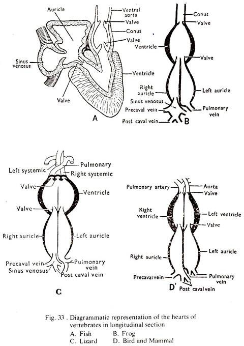
Learn about the comparison of heart in various vertebrates.
Comparison: Vertebrates # Lates:
1. The heart is enclosed within the pericardium and is placed on the ventral side below the oesophagus. It consists of 3 chambers, a sinus venosus, an a uricle and ventricle.
2. The sinus vernosus is a thin walled chamber situated dorsal to the auricle. It receives deoxygenated blood from the body, through a cuvierian duct from each side. The sinus venosus opens into the auricle by a sinuauricular aperture.
3. The auricle is a large thin-walled chamber, ventral to the sinus venosus. It opens into the ventricle by an auriculo-ventricular aperture.
ADVERTISEMENTS:
4. The ventricle is a small thick-walled chamber, ventral to the auricle. Anteriorly, it gives off a stout median vessel, the ventral aorta, the base of which in incorporated in the pericardial cavity and is swollen to form the bulbus aorta. The ventral aorta runs forward and sends blood.
5. Valves. All the apertures are guarded by valves which prevent the back-flow of blood.
6. Mechanism of circulation. Deoxygenated blood from the body comes to the sinus venosus through a pair of cuvierian ducts. Blood from the sinus passes to the auricle through the sinuauricular aperture, and from there to the ventricle through the auriculoventricular aperture.
From the ventricle blood passes to the gills through the ventral aorta. Alternate relaxation and contraction of the heart is going on and the sum total of the two is known as a heartbeat. The heart contains deoxygenated blood and the circulation in fish is known as single circulation.
Comparison: Vertebrates # Bufo:
ADVERTISEMENTS:
1. The heart is enclosed within the pericardium and is placed ventrally in the thoracic cavity. It consists of five chambers, a sinus venosus, the right and left auricles, a ventricle and a conus arteriosus.
2. The sinus venosus is a thin walled chamber placed on the dorsal side of the heart. It receives deoxygenated blood from the three venae cavae and opens into the right auricle by a sinuauricular aperture.
3. The two auricles are thin-walled and placed in front of the ventricle. The two auricles are separated by an interauricular septum. The right auricle is larger and receives deoxygenated blood from the sinus. The left auricle receives oxygenated blood from the lungs through the pulmonary veins. The two auricles open into the ventricle by a common auriculoventricular aperture.
4. The ventricle is a thick-walled chamber with the pointed end directed backward. It has a small cavity and a thick muscular wall. The internal Wall of the Ventricle is thrown into muscular ridges known as columnae carane.
ADVERTISEMENTS:
(a) From the anteroventral right corner of the ventricle arises a stot tube, the conus arteriosus which is oriented obliquely towards the left side. The conus has a spiral longitudinal valve within it. The conus runs forward and opens into a truncus arteriosus, which bifurcates and each branch is again divided into three arches by two partitions.
5. Valves. All the apertures are guarded by valves which prevent the backflow of blood. Three semilunar valves are present at the base of the conus.
6. Mechanism of Circulation. Deoxygenated blood from the body comes to the sinus venosus through three main veins and from there to the right auricle through the sinuauricular aperture. The left auricle receives oxygenated blood from the lungs. The two auricles contract and the blood is driven to the ventricle through the common auriculoventricular aperture.
A somewhat mixture of oxygenated and deoxygenated blood takes place in the ventricle. The ventricle contracts and the blood is driven into the conus. The spiral valve in the conus guides the blood into the truncus and from there to the arterial arches.
Comparison: Vertebrates # Calotes:
1. The heart is enclosed within the pericardium and is placed ventrally in the thoracic cavity. It consists of four chamber, a sinus venosus, the right and left auricles, and an incompletely divided ventricle.
2. The sinus venosus is a thin-walled chamber place on the dorsal surface of the heart into which open the three venae cave. The sinus venosus opens into the right auricle by a sinuauricular aperture.
3. The two auricles are thin-walled and placed in front of the ventricle. The inner surfaces of the auricles are provided with muscular ridges. The right auricle receives deoxygenated blood from the sinus venosus and the left auricle receives oxygenated blood from the lungs through pulmonary veins. The two auricles open into the ventricle by a common auriculoventricular aperture divided into two by the interauricular septum.
4. The ventricle is a thick-walled structure with a small cavity divided into two parts by an incomplete muscular partition.
(a) From the right portion of the ventricular cavity (cavum ventralee) arises the pulmonary artery and from the left and right side of the cavum dorsale arise the right and left systemic arches respectively. The carotid arch arises from the right systemic arch.
ADVERTISEMENTS:
5. Valves. All the apertures are guarded by valves which prevent the backflow of blood. The auriculoventricular aperture and the openings of the arches are guarded by semilunar valves.
6. Mechanism of circulation. Deoxygenated blood from the body comes to the sinus venosus through three main veins and from there to the right auricle through the sinuauricular aperture. The left auricle receives oxygenated blood from the lungs.
The two auricles contract and the deoxygenated blood from the right auricle tends to run more to the right hand cavity and the oxygenated blood from the left auricle to the left hand cavity of the ventricle. The ventricle contracts and the incomplete partition separates the two cavities of the ventricle.
The pulmonary arch being placed on the right side receives deoxygenated blood which goes to the lungs for oxidation. The left systemic arch being placed on the middle receives mixed blood which goes to the posterior part of the body. The right systemic being placed on the left side receives almost oxygenated blood which goes to the anterior end of the body, the region of the highest activity.
Comparison: Vertebrates # Columba:
1. The heart is enclosed within the pericardium and is placed ventrally in the thoracic cavity. It consists of four chambers, the right and left auricles and the right and left ventricles. The right ventricle partly encircles the left.
2. The auricles are comparatively thick-walled and placed in front of the ventricles. The inner surfaces of the auricles are provided with muscular ridges. The right auricle receives deoxygenated blood from the body through the two pre-cavals and one post-caval. The left auricle receives oxygenated blood from the lungs through four pulmonary veins. The right and left auricles open into the right and left ventricles respectively through auriculo ventricular apertures.
3. The ventricles are thick-walled structures with small cavities.
(a) From the right ventricle arises the pulmonatry artery and from the left ventricle arises the aortich.
4. All the apertures are guarded by valves which prevent the back-flow of blood. The valve guarding the right auriculoventricular aperture, is a large muscular fold found in no other vertebrates. The opening of the aorta is guarded by semilunar valves.
5. Mechanism of circulation. Deoxygenated blood from the body comes to the right auricle through three main veins. The left auricle receives oxygenated blood from the lungs. The auricles contract and the deoxygenated blood from the right auricle and oxygenated blood from the left auricle pass to the right and left ventricles through the auriculoventricular apertures.
These are completely separated structures and no mixing of blood can take place at any point. The ventricles contract and deoxygenated blood from the right ventricle goes to the lungs through the pulmonary artery. Oxygenated blood from the left ventricle goes to the body through the right aortic arch.
Comparison: Vertebrates # Cavia:
1. The heart is enclosed within the pericardium and is placed ventrally in the thoracic cavity. It consists of four chambers, the right and left auricles and the right and left ventricles. The pericardium consists of two layers. The right ventricle partly encircles the left.
2. The auricles are comparatively thick-walled, placed in front of the ventricles. The inner surfaces of the auricles are provided with muscular ridges. The interauricular septum bears a fossa ovalis. The right auricle receives deoxygenated blood from the body through the anterior and posterior venae cavae.
The left auricle receives oxygenated blood from the lungs through four pulmonary veins. Both the auricles are provided with auricular appendices. The right and left auricles open into the right and left ventricles respectively through auriculoventricular apertures.
3. The ventricles are thick-walled structures with small cavities.
(a) From the right ventricle arises the pulmontary artery and from the left ventricle arises the aortic arch.
4. All the apertures are guarded by valves which prevent the back-flow of blood. The right auriculoventricular aperture is guarded by tricuspid and the left by bicuspid valves. The openings of both the pulmonary artery and the aorta are guarded by three semilunar valves.
6. Mechanism of circulation. Deoxygenated blood from the body comes to the right auricle through two venae cavae. The left auricle revceives oxygenated blood from the lungs. The auricles contract and the deoxygenated blood from the right and the oxygenated blood from the left pass to the right and the left ventricles through the auriculoventricular apertures.
These are completely separated structures and no mixing of blood can take place at any point. The ventricles contract and deoxygenated blood from the right ventricle goes to the lungs through the pulmonary artery. Oxygenated blood from the left ventricles goes to the body through the left aortic arch.
