In this article we will discuss about the Development of Brain in Chick:- 1. Introduction to Development of Brain in Chick 2. Formation of Three-Part Brain in Chick 3. Formation of Five-Part Brain 4. Formation of Flexures 5. Out-pocketing’s of the Brain 6. Cavity of the Primitive Five-Part Brain 7. Embryonic Induction in Brain Development.
Contents:
- Introduction to Development of Brain in Chick
- Formation of Three-Part Brain in Chick
- Formation of Five-Part Brain in Chick
- Formation of Flexures in Chick
- Out-pocketing’s of the Brain in Chick
- Cavity of the Primitive Five-Part Brain in Chick
- Embryonic Induction in Brain Development of Chick
1. Introduction to Development of Brain in Chick:
The neural tube formed from the neural ectoderm is located dorsally in the median plain of the embryo and forms the basis for the central nervous system. Before going into the development of brain a brief idea about the formation of neural tube in chick is dealt with.
ADVERTISEMENTS:
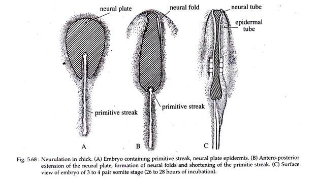
During gastrulation of chick a primitive streak with Hensen’s node is formed. In the 24 hours chick-embryo, as epiboly of ectodermal cells is taking place at the surface of the embryo, it brings about an elongation of the presumptive neural ectodermal cells above the notochordal area (Fig. 5.68).
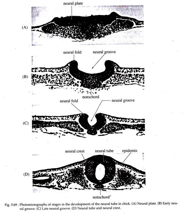
As gastrulation comes to an end, the single-layered neural ectoderm rapidly becomes thick and stratified to form the neural plate. Starting from the anterior towards the posterior side, the neural plate gradually sinks down and its margins becomes elevated resulting in the formation of a neural groove bounded by the neural folds (Fig. 5.69).
The groove continues to deepen and the neural folds meet above it, converting the original plate into a neural tube. The meeting of the neural folds is accompanied by the meeting of the epidermal ectoderm, which results in detaching the neural tube from the overlying ectoderm.
ADVERTISEMENTS:
The primitive neural tube is composed of two major regions — the future brain region at its anterior end and the rudiments of the spinal cord at the posterior end. The brain, thus, can be defined simply as the anterior part of the neural tube lying within the cranium. However, its morphogenetic history is one of increasing complexity of both gross anatomical form and internal organization.
The brain of Amphioxus hardly differs architecturally from the spinal cord. However, in amphibians to mammals, the prospective brain differs from the spinal cord behind. Therefore, in chick the fore part of the neural tube is considerably wider than that of the spinal tube.
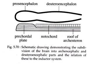
As the neural folds are about to meet (at the time of formation of the neural tube), it is possible to distinguish two subdivisions in the forming brain. The primordial brain tube is initially divided into an anterior pros-encephalon (archencephalon) and posterior deuteroencephalon (Fig. 5.70).
The regionally specific induction mechanisms influence the formation of the neural tube. Thus, the pros encephalon is induced by prechordal plate mesoderm and the deuteroencephalon by the anterior portion of the notochordal mesoderm, both lying beneath the primordial brain.
2. Formation of Three-Part Brain in Chick:
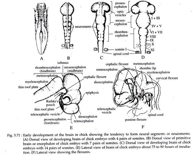
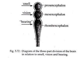
The deuteroencephalon undergoes a constriction termed the isthmus (Fig. 5.71E), that divides it into a mesencephalon (mid brain) behind the pros encephalon (fore brain), and a rhomb encephalon (hind brain) in front of the spinal tube (Fig. 5.72). Thus, the brain is said to pass through a three-part stage.
The distinction between these three divisions rests more on functional than anatomical ground. Accordingly, the pros encephalon is associated with the sense of smell, the mesencephalon with vision and the rhomb encephalon with hearing.
The anterior end of the neural tube during its early stages of development, tends to form primitive segments called neuromeres. These neuromeres fuse as they contribute to the primitive brain regions (Fig. 5.71A-D).
3. Formation of Five-Part Brain in Chick:
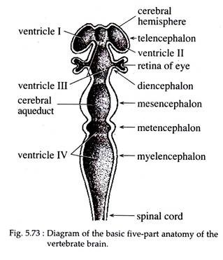
Further embryonic modifications result in a subdivision of two of the three primary brain regions at about 75 to 80 hours of incubation. The pros encephalon is divided into anterior telencephalon and a posterior diencephalon. The mesencephalon remains intact. The rhomb encephalon is also divided into two, forming an anterior metencephalon and a posterior medulla or myelencephalon (Fig. 5.73).
ADVERTISEMENTS:
The five divisions of the brain are arranged generally in a straight line in case of lower vertebrates. However, in case of chick and other higher vertebrates, the brain tube tends to bend upon itself at an early period of its embryogeny, by formation of flexures.
4. Formation of Flexures in Chick:
Flexures are formed at three regions to create sharp angles in the long axis of the embryonic brain. These are accompanied by a more general flexing of the entire head. At the time of the primordial brain tube formation a primary neural flexure occurs at the mesencephalon region called the cephalic flexure.
The cephalic flexure is more pronounced in birds. It sharply bends the fore- brain to the ventral side (Fig. 5.71E). Soon thereafter, a second flexure called cervical flexure develops at the caudal portion of the myelencephalon and the anterior part of the spinal cord.
It bends the entire brain region of chick ventrally (Fig. 5.71F) and is pronounced in birds like that of other amniotes. A third flexure or pontine flexure appears later at the mid-region of the rhomb encephalon, in the area between myelencephalon and metencephalon.
It counter acts the effect of the other two flexures by bending the brain in the opposite direction (Fig. 5.71 F), that is dorsally. The pontine flexure is found only in the higher vertebrates.
5. Out-pocketing’s of the Brain in Chick:
As the five primary subdivisions of the brain are established, its wall exhibits a number of local out-pocketing’s. The telencephalon gives rise to two lateral pouches, the telencephalic vesicles (Fig. 5.71 E). The telencephalic vesicles represent the rudiments of the cerebral lobes. The diencephalon on the other hand forms four or five evaginations.
These are — a mid-dorsal evagination, the epiphysis (rudiments of the pineal body); a second mid-dorsal evagination occurs in front of the epiphysis called paraphysis; two ventro-lateral outgrowths, the optic vesicles (Fig. 5.71 C and D) and the infundibulum arising from a mid-ventral evagination.
The Rathke’s pouch (Fig. 5.71E) arising from the stomodaeum unites with the infundibulum and ultimately differentiates into the anterior lobe of the pituitary body.
From the mesencephalic roof or tectum dorsal swellings appear. In birds like other amniotes, four swellings arise in the tectum, the corpora quadrigemina. From the roof of the metencephalon there arise two cerebellar outpushings.
These out-pocketing’s are accompanied by continued cell multiplications. The differentiating neuro-blasts undergo localized aggregations that bring about thickening of gray and white matter and a variety of folds, fissures and invaginations. The roof of the diencephalon and myelencephalon are thin, highly vascular and are devoid of nervous tissue.
6. Cavity of the Primitive Five-Part Brain in Chick:
The brain as well as the spinal cord are hollow structures and this cavity is generally called the neural cavity or neurocoel (Fig. 5.73). The neurocoel are fluid-filled cavities. The cavities of the telencephalic vesicles extend into the cerebral hemispheres (cerebral lobes) and are known as the lateral ventricles or the first and second ventricles.
These ventricles communicate with the lumen of the middle of the telencephalon and that of the diencephalon, which together form the third ventricle.
The cavity of the mesencephalon is designated as the cerebral aqueduct or aqueduct of Sylvius. It becomes a narrow passage way and greatly reduced in chick as well as in other amniotes. The cavity of the rhomb encephalon is termed as the fourth ventricle. The cerebral aqueduct connects the third ventricle in front with that of the fourth.
In the roof of the third and fourth ventricles, small blood vessels from the pia mater form freely branching groups of vessels and are called the anterior and posterior choroid plexus. These plexus, plus those that form the walls of the lateral ventricles secrete cerebrospinal fluid that bathes the tissues of the brain and spinal cord.
7. Embryonic Induction in Brain Development of Chick:
Activation phase of induction results primarily in the formation of neural tube. The normal prospect of the neural tube is its differentiation into four general regions. The most anterior of it will become the fore brain.
Subsequent regions will provide the mid brain and hind brain. This is then followed by the prospective spinal cord. Finally there is an area that fails to produce neural structures at all but contributes to the mesodermal composition of the tail.
The question that may arise is, how the pattern of regions appearing in the neural tube may be related to a pattern present in the inducing mesoderm? This is indicated by two sets of facts. First, in the anatomical fact the notochord terminates anteriorly at approximately the junction between the fore- and mid-brain, the remaining forward distance being occupied by the prechordal mesoderm or plate.
This prechordal plate may thus be considered the inductor of the fore brain and the notochord the inductor of the regions behind. The second fact is that different levels of the mesoderm have unlike inductive capacities. If different regions of the prospective chorda mesoderm from anterior to posterior are cut out and grafted into young gastrula, it is found that they induce characteristically different regions of the neural tube.
Immediately after the neural plate is established, this neural primordium itself acts as a secondary inductor. Some examples are induction of sensory structures (lens of the eye etc.), the otic vesicle (inner ear) and the series of ectodermal placodes. The brain and the spinal cord later, also induces the formation of bony structure that protects them.
