In this article we will discuss about the Gametogenesis:- 1. Meaning of Gametogenesis 2. Origin of Gametogenesis (Primordial Germ Cells).
Meaning of Gametogenesis:
There are two types of reproductive cells called gametes — the spermatozoa in males and ova in females. These reproductive sex cells and the cells that give rise to them are called germplasm.
On the other hand, the cells of the body that do not take part in the production of the gametes are called somatic cells and are collectively called somatoplasm. The germplasms are the ones, constituting the hereditary material that is passed on from one generation to the other, of a species.
Gametogenesis refers to the processes by which germplasm is converted into highly specialized sex cells that are capable of uniting during fertilization and in producing a new individual. It includes spermatogenesis in males and oogenesis in females. In both the cases the diploid germplasm is converted into the haploid male (spermatozoon) and female (ovum) sex cells.
ADVERTISEMENTS:
Generally gametogenesis is divided into four major phases. These are:
(a) Origin of the germ cells (called primordial germ cells or PGC) and their migration to the gonads,
(b) Multiplication of the germ cells in the gonads by mitosis,
(c) Reduction in the number of chromosomes to half by meiosis and
ADVERTISEMENTS:
(d) The final stages of maturation and differentiation of the gametes into either spermatozoa or ova. The last three phases are described under spermatogenesis and oogenesis.
Origin of Gametogenesis (Primordial Germ Cells):
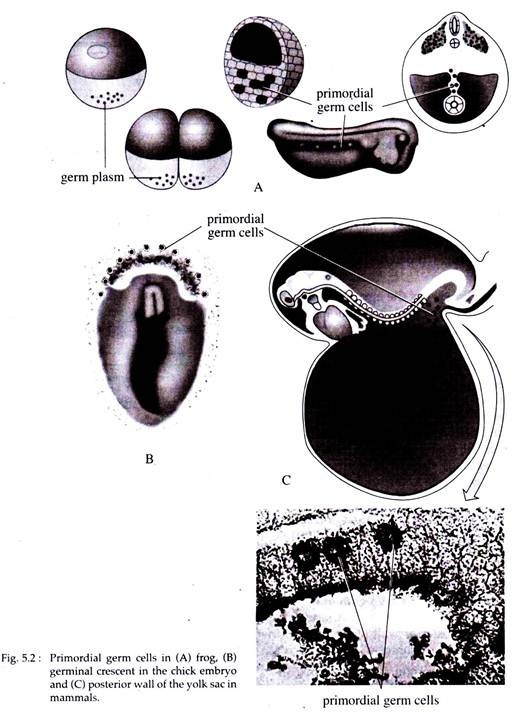
The first step in gametogenesis involves the formation of primordial germ cells (PGCs) and their travelling to the genital ridge as the gonad is forming. In most animal species (in frogs and a number of invertebrate species) the determination of the PGCs is brought about by the localization of specific proteins and mRNA in the cytoplasm of the early embryo.
These cytoplasmic components are said to be the germplasm. The germplasm in frogs and a number of invertebrate species, is recognized sometimes as regions in the vegital pole cytoplasm or as specific cells during the cleavage stages (Fig. 5.2A).
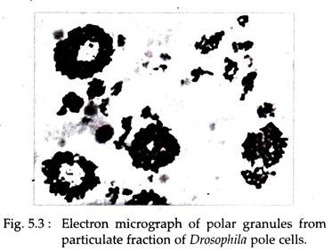
In Drosophila, however, the germ cells become distinct at the posterior pole of the zygote, about 90 minutes after fertilization and several hours before cellularization of the rest of the embryo. This posterior cytoplasm, the so-called pole plasm, is made of a complex collection of mitochondria, fibrils and polar granules (Fig. 5.3).
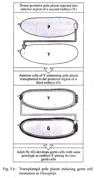
That this pole plasm is responsible for the development of germ cells can be demonstrated by two experiments:
a. If the posterior end of the egg is subjected to ultraviolet (UV) irradiation (it destroys the pole plasm activity), no germ cells develop.
b. If pole plasm of an egg is transferred to the anterior pole of a second embryo, the nucleuses that become surrounded by the pole plasm get specified as germ cell. If these germ cells are transplanted into the future genital region of a third embryo, they develop as functional germ cells with same genotype as that of the second embryo (Fig. 5.4).
Primordial germ cells of reptiles, birds and mammals arise in the epiblast of early embryo and take up temporary position in the extra embryonic tissues. In birds they are located in the germinal crescent, which is present beyond the future head region of the embryo (Fig. 5.2B). In mammals PGCs are recognized in the posterior Wall of the yolk sac at the junction of the allantosis (Fig. 5.2C).
Regardless of their origin, PGCs are easily recognizable because of their large size, clear cytoplasm and by certain histochemical characteristics. The histochemical characteristic of PGCs in mammals is high alkaline phosphatase activity, while in birds it is high in glycogen content.
Primordial Germ Cell Migration:
At the time of recognition of PGCs in embryos, the gonads are either poorly developed or not developed at all.
ADVERTISEMENTS:
Later, PGCs in vertebrates migrate to the gonads mainly by two mechanisms:
a. In birds and reptiles, PGCs migrate through the wall of local blood vessels and enter the circulation. Through the circulation they recognize the blood vessels of the gonads, penetrate through their wall and settle down in the gonads.’
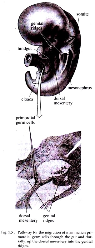
b. Mammalian PGCs migrate around the wall of the posterior gut, then through the dorsal mesentery they, reach the gonads (Fig. 5.5). Studies have revealed that the number of primordial germ cells increases during their migration to the gonads. The number of PGCs in mouse rises from less than 100 to about 5,000 during the period of migration.


