In this article we will discuss about:- 1. Structure of Liver Fluke 2. Reproductive Organs of Liver Fluke 3. Fertilization and Development 4. Life History 5. Effect of Parasitism.
Structure of Liver Fluke:
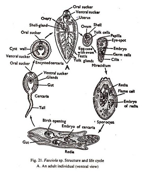
1. Body of liver fluke is soft, flattened, leaf-like with a triangular head lobe (Fig. 21 A). It is brown to pale-grey in colour and measures 2.15-3 cm x 1.2-1.5 cm. The body is covered with a cuticle, the greater portion of which bears minute spines.
2. The mouth of liver fluke is anterior and terminal, surrounded by the oral sucker.
3. The posterior sucker is ventral and behind the mouth.
ADVERTISEMENTS:
4. The genital aperture is placed in between the two suckers.
5. The excretory pore is median and posterior terminal in liver fluke.
Reproductive Organs of Liver Fluke:
1. Liver fluke is hermaphrodite and bears organs of both the sexes.
2. The male organs consist of a paired, much branched testes, a pair of vasa deferentia, a common vesicula seminalis, and an ejaculatory duct opening in the male aperture in the cirrus.
ADVERTISEMENTS:
3. The female organs of liver fluke consist of a single branched ovary, an oviduct, a uterus, an ootype, vitelline glands and their ducts and shell glands. The uterus opens in the cirrus.
4. A small cavity, the genital atrium bearing the external aperture of male and female duct is formed when the cirrus is withdrawn.
Fertilization and Development in Liver Fluke:
1. The eggs of liver fluke are large ovoidal structures with brown colour due to the presence of bile pigment. On the average, they measure 40-80yum (microns). The eggs are fertilized in the uterus and self or cross fertilization may occui.
2. The zygote receives yolk and a chitinoid covering in the ootype, remains in the uterus for a short period and is discharged. Passing down the bile duct, the zygote reaches the intestine of the host and passes to the exterior with the faeces.
ADVERTISEMENTS:
3. Active development starts and after 2-3 weeks the miracidiurn larva hatches out of the egg.
Life History of Liver Fluke:
Miracidiurn larva:
(a) The body of liver fluke is conical with a triangular head lobe and is covered with vibratile cilia.
(b) A pair of crossed eye spots are present anteriorly.
(c) An imperfectly developed intestine and a pair of flame cells opening to the exterior are present.
(d) The rest of the interior is filled up with a mass of germ cells.
4. The miracidium swims actively in water or moves on damp herbage and can survive only if it reaches an amphibian snail, the other host, approximately within 8 hours-time.
5. The embryo bores into the snail and comes to the pulmonary sac or other organs.
6. The ciliated ectoderm is lost, it grows to an elongated sac and forms the sporocyst.
Sporocyst:
(a) Its wall is formed by a single layer of cells.
(b) The flame cells and remnants of eye spots are present.
(c) The internal cavity contains germ cells.
7. The germ cells of the sporocyst behave like parthenogenetic ova. Each cell divides to produce blastula, gastrula and finally a form of larva, the redia.
8. Five to eight rediae are usually formed in each sporocyst.
Redia:
(a) The body is cylindrical, with a circular ridge near the anterior end and a pair of short processes near the posterior end.
(b) The mouth leads into pharynx and a sac-like intestine is present.
(c) A system of excretory vessel is present.
(d) Germ cells are present in the internal cavity.
(e) A birth opening is situated anteriorly near the circular edge.
9. From the germ cells in the redia, 14-20 cercariae develop in each redia. Cercariae are produced if the season is summer but redia gives rise to a fresh generation of 8-12 rediae if the season is winter.
10. The cercaria escapes through the birth opening of the redia.
Cere aria:
(a) The body is oval with a long tail.
(b) The anterior and posterior suckers, the mouth, pharynx and a bifid intestine are present.
(c) The gonads, glands, etc., begin to appear.
11. The cercaria forces its way out of the snail, loses its tail, becomes encysted and remains attached to blades of grass or other herbage.
12. The encysted cercaria known as metacercaria, is taken by the host with the grass and the young fluke on escape, may reach the liver through bile ducts or hepatic portal vein, and grows rapidly to reach the adult stage. The eggs come out with the faeces about 3 to 4 months (incubation period) after infection.
Effect of Parasitism on Liver Fluke:
1. On host:
The attack of liver fluke causes ‘liver-rot’, which is disastrous to the host and death has been recorded in most cases of liver-rot. Jaundice and adenomata have also been reported.
2. On parasite:
Due to parasitic life, considerable degeneration of the vegetative organs has taken place in Fasciola. The reproductive organs are more developed. On the other hand a single fluke may produce about 50,000 eggs. Twice in its life cycle, the young embryos are exposed to the environment and the cycle which is already full of risks becomes more risky.
To compensate the huge loss during its perilious journey from host to host further multiplication by asexual means has appeared, in addition to the already accentuated rate of multiplication.
