Learn about the comparison of respiratory system in various Arthropods.
Comparison: Arthropod # Macrobrachium:
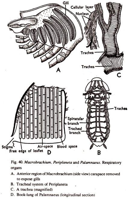
1. The respiratory organs consist of the lining membrane of the bran- chiostegite, three pairs of epipodites and eight pairs of gills. The gills are lodged in gill chambers, which communicate with the exterior along its anterior, posterior and ventral borders.
ADVERTISEMENTS:
3. Each gill consists of a long, narrow rachis supporting two rows of rhomboidal gill-plates diverging from each other at right angles to the elongated axis. The gill-plates are larger in size in the middle but smaller towards the ends. Each gill-plate is made up of a double layer of cuticle with a single layer of cells in between. The gill is attached to the body about the middle of its length, and is highly vascular.
4. The axis of the gill is roughly triangular in cross section. Three longitudinal canals, two laterals and one median, run along the axis. The laterals are connected with each other by transverse channels and also with the median canal by marginal channels. Blood enters through the transverse channels and traverses other channels. Gaseous exchange takes places in the gill filaments.
5. The lining membrane of the branchio-stegite and the epipodites of the 3 maxillipeds are highly vascular and aid in the process of respiration.
6. The movements of scaphognathites maintain a constant backward to forward water current in the gill-chambers. The blood in the respiratory organs gives up CO2 and absorbs O2.
Comparison: Arthropod # Periplaneta:
ADVERTISEMENTS:
1. The respiratory organs consist of tracheae or air tubes and their branches or tracheoles which are directly communicated with the exterior.
2. The tracheae open to the exterior by ten pairs of apertures, the spiracles or stigmata. Two are placed on each side of the thorax; one between pro- and meso- thorax and the other between meso- and meta- thorax. Eight occur on each side of the abdomen between terga and sterna of the first eight segments.
The stigmata are guarded by hairs, etc., to prevent the entrance of harmful particles. Each spiracle is controlled by muscular valves. From each spiracle leads inward a small tube which ends in a stout trachea and all of them are connected by longitudinal and transverse tracheae.
3. There are two lateral longitudinal tracheal trunks, one on each side of the body. They are connected by several transverse tracheae, and the tracheae form a system of intercommunicating network. Each trachea divides and subdivides into numerous branches and the ultimate, very fine branches entering the organs are called tracheoles. The tracheoles end in tissues. The wall of the trachea is strengthened by chitinoid lining in the form of spiral threads.
ADVERTISEMENTS:
6. The alternate expansion and contraction of the abdomen brings inhalation and exhalation of air in the tracheal system. The opening and closing of the spiracles are also dependent on the CO2 concentration of the inhaled air. The walls of the tracheoles allow gaseous exchange and diffusion takes place between the cell sap and the lumen of the tube. The tissues get direct supply of oxygen.
Comparison: Arthropod # Palamnaeus:
1. The respiratory organs consist of four pairs of book- lungs, which open in four pairs of stigmata.
2. The book-lungs open to the exterior by four pairs of apertures, the stigmata. Sternum of each of the 3rd, 4th, 5th and 6th preabdominal segments bears a pair of stigmata on its lateral sides. These are narrow oblique slits. Each stigmata opens directly into a book-lung and they are not interconnected.
3. Each book-lung is a compressed sac lined with a thin cuticle. The lining membrane is folded up into numerous delicate laminae lying parallel to one another like the pages of a book, roughly 130-150 in number.
4. Deoxygenated blood flows through the narrow space in each lamina, separated from the air only by the membranous walls of the lamina.
6. The alternate expansion and contraction of the abdomen brings inhalation and exhalation of air in the book-lungs. Gaseous exchange takes place between the blood in the laminae and the air in between the laminae.
