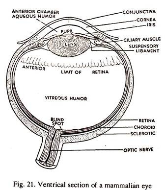
In this article we will discuss about the structure of mammalian eye with the help of diagram. Also learn about its functions and accommodation.
The wall of the eye ball has got three concentric layers.
1. The outermost layer of the eye ball is known as tunica fibrosa, skeletal in function, and maintains the rigidity of the eye ball. It consists of sclera and choroid. The sclera is opaque, and is supplied by optic nerve and blood vessels, but in front, it is replaced by a transparent layer called cornea. A thin mucous membrane, in continuation with the eye lids, is present in front of the cornea. This is the conjunctiva.
2. The layer beneath the sclera is called tunica vasculosa. It is composed of choroid, ciliary body and iris, rich in blood vessels and pigment cells. Behind the cornea, the choroid coat is a pigmented disc and is known as iris. A large aperture, the pupil is present at the centre of the iris. The iris is provided with ciliary muscles by which the pupil can be contracted or dilated.
ADVERTISEMENTS:
3. The third or innermost coat of the eye is retina. It contains rods and cones which receive the visual impulses. The retina acts as a sensitive screen on which the images of objects are focussed. The retinal layer is highly innervated by the optic nerve. The point where the optic nerve enters the retina is known as the blind spot.
4. The lens is biconvex and situated just behind the pupil.
5. The space between the cornea and the lens is the anterior chamber and it is filled up with a thin watery fluid, the aqueous humour.
6. The space between the lens and the retina is the posterior chamber and is filled up with a jelly-like substance, the vitreous humour.
Functions of Mammalian Eye:
Light rays enter the eye ball through the transparent cornea and are focussed by the lens on the retina. An inverted image of the object is formed on the retina and this is conveyed to the visual centres of the optic lobes in the brain by the optic nerves. The inverted image is corrected and a direct image is formed.
Accommodation of Mammalian Eye:
Normally, the eye is focussed for distant vision. It can be adjusted for near vision by increasing the convexity of the lens. Due to the tendency of the eye ball to remain globular normally, there is a pull on the lens which keeps it flat.
Contraction of the ciliary muscles releases this pull on the lens and the elastic lens becomes more convex. The divergent rays from the near object are thus focussed to form a clear image on the retina.
